The structure in solution of the b domain of protein disulfide isomerase.
Kemmink, J., Dijkstra, K., Mariani, M., Scheek, R.M., Penka, E., Nilges, M., Darby, N.J.(1999) J Biomol NMR 13: 357-368
- PubMed: 10383197
- DOI: https://doi.org/10.1023/a:1008341820489
- Primary Citation of Related Structures:
1BJX, 2BJX - PubMed Abstract:
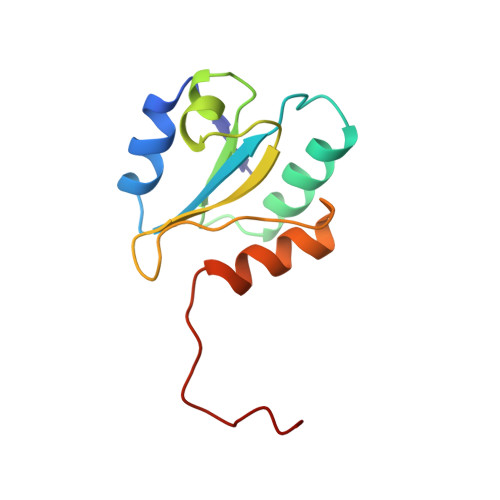
Protein disulfide isomerase (PDI) is a multifunctional protein of the endoplasmic reticulum, which catalyzes the formation, breakage and rearrangement of disulfide bonds during protein folding. It consists of four domains designated a, b, b and a. Both a and a domains contains an active site with the sequence motif -Cys-Gly-His-Cys-involved directly in thiol-disulfide exchange reactions. As expected these domains have structures very similar to the ubiquitous redox protein thioredoxin. A low-resolution NMR structure of the b domain revealed that this domain adopts a fold similar to the PDI a domain and thioredoxin [Kemmink, J., Darby, N.J., Dijkstra, K., Nilges, M. and Creighton, T.E. (1997) Curr. Biol. 7, 239-245]. A refined ensemble of solution structures based on the input of 1865 structural restraints shows that the structure of PDI b is well defined throughout the complete protein except for about 10 residues at the C-terminus of the sequence. 15N relaxation data show that these residues are disordered and not part of this structural domain. Therefore the domain boundaries of PDI can now be fixed with reasonable precision. Structural comparison of the PDI b domain with thioredoxin and PDI a reveals several features important for thiol-disulfide exchange activity.
Organizational Affiliation:
European Molecular Biology Laboratory 9EMBL), Heidelberg, Germany. johan.kemmink@oulu.fi