2BGR

Crystal structure of HIV-1 Tat derived nonapeptides Tat(1-9) bound to the active site of Dipeptidyl peptidase IV (CD26)


Experimental Data Snapshot

  • Method: X-RAY DIFFRACTION
  • Resolution: 2.00 Å
  • R-Value Free: 0.203 
  • R-Value Work: 0.160 
  • R-Value Observed: 0.160 

wwPDB Validation   3D Report Full Report


This is version 2.1 of the entry. See complete history


Literature

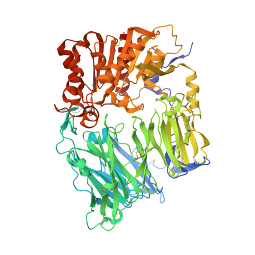
Crystal Structures of HIV-1 Tat-Derived Nonapeptides Tat-(1-9) and Trp2-Tat-(1-9) Bound to the Active Site of Dipeptidyl-Peptidase Iv (Cd26)

Weihofen, W.A.Liu, J.Reutter, W.Saenger, W.Fan, H.

(2005) J Biol Chem 280: 14911

  • DOI: https://doi.org/10.1074/jbc.M413400200
  • Primary Citation of Related Structures:  
    2BGN, 2BGR

  • PubMed Abstract: 

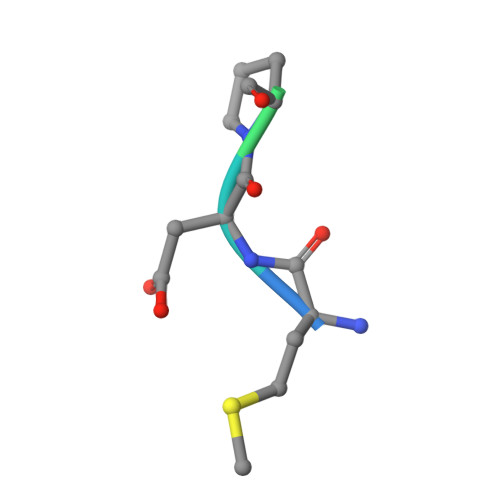
    CD26 or dipeptidyl-peptidase IV (DPPIV) is engaged in immune functions by co-stimulatory effects on activation and proliferation of T lymphocytes, binding to adenosine deaminase, and regulation of various chemokines and cytokines. DPPIV peptidase activity is inhibited by both Tat protein from human immunodeficiency virus (HIV)-1 and its N-terminal nonapeptide Tat-(1-9) with amino acid sequence MDPVDPNIE, suggesting that DPPIV mediates immunosuppressive effects of Tat protein. The 2.0- and 3.15-A resolution crystal structures of the binary complex between human DPPIV and nonapeptide Tat-(1-9) and the ternary complex between the variant MWPVDPNIE, called Trp(2)-Tat-(1-9), and DPPIV bound to adenosine deaminase show that Tat-(1-9) and Trp(2)-Tat-(1-9) are located in the active site of DPPIV. The interaction pattern of DPPIV with Trp(2)-Tat-(1-9) is tighter than that with Tat-(1-9), in agreement with inhibition constants (K(i)) of 2 x 10(-6) and 250 x 10(-6) m, respectively. Both peptides cannot be cleaved by DPPIV because the binding pockets of the N-terminal 2 residues are interchanged compared with natural substrates: the N-terminal methionine occupies the hydrophobic S1 pocket of DPPIV that normally accounts for substrate specificity by binding the penultimate residue. Because the N-terminal sequence of the thromboxane A2 receptor resembles the Trp(2)-Tat-(1-9) peptide, a possible interaction with DPPIV is postulated.


  • Organizational Affiliation

    Institut für Chemie/Kristallographie, Freie Universität Berlin, Takustrasse 6, D-14195 Berlin, Germany.


Macromolecules
Find similar proteins by:  (by identity cutoff)  |  3D Structure
Entity ID: 1
MoleculeChains Sequence LengthOrganismDetailsImage
DIPEPTIDYL PEPTIDASE IV
A, B
738Homo sapiensMutation(s): 0 
EC: 3.4.14.5
UniProt & NIH Common Fund Data Resources
Find proteins for P27487 (Homo sapiens)
Explore P27487 
Go to UniProtKB:  P27487
PHAROS:  P27487
GTEx:  ENSG00000197635 
Entity Groups  
Sequence Clusters30% Identity50% Identity70% Identity90% Identity95% Identity100% Identity
UniProt GroupP27487
Sequence Annotations
Expand
  • Reference Sequence

Find similar proteins by:  Sequence   |   3D Structure  

Entity ID: 2
MoleculeChains Sequence LengthOrganismDetailsImage
HIV-1 TAT PROTEIN DERIVED N-TERMINAL NONAPEPTIDEC [auth Y],
D [auth Z]
9Human immunodeficiency virus 1Mutation(s): 0 
UniProt
Find proteins for P12506 (Human immunodeficiency virus type 1 group M subtype D (isolate Z2/CDC-Z34))
Explore P12506 
Go to UniProtKB:  P12506
Entity Groups  
UniProt GroupP12506
Sequence Annotations
Expand
  • Reference Sequence
Oligosaccharides

Help

Entity ID: 3
MoleculeChains Length2D Diagram Glycosylation3D Interactions
2-acetamido-2-deoxy-beta-D-glucopyranose-(1-4)-[alpha-L-fucopyranose-(1-6)]2-acetamido-2-deoxy-beta-D-glucopyranoseE [auth C]3N-Glycosylation
Glycosylation Resources
GlyTouCan:  G21290RB
GlyCosmos:  G21290RB
GlyGen:  G21290RB
Entity ID: 4
MoleculeChains Length2D Diagram Glycosylation3D Interactions
2-acetamido-2-deoxy-beta-D-glucopyranose-(1-4)-2-acetamido-2-deoxy-beta-D-glucopyranose
F [auth D],
H [auth F],
I [auth G],
K [auth I],
M [auth K],
F [auth D],
H [auth F],
I [auth G],
K [auth I],
M [auth K],
N [auth L]
2N-Glycosylation
Glycosylation Resources
GlyTouCan:  G42666HT
GlyCosmos:  G42666HT
GlyGen:  G42666HT
Entity ID: 5
MoleculeChains Length2D Diagram Glycosylation3D Interactions
beta-D-mannopyranose-(1-4)-2-acetamido-2-deoxy-beta-D-glucopyranose-(1-4)-2-acetamido-2-deoxy-beta-D-glucopyranoseG [auth E],
L [auth J]
3N-Glycosylation
Glycosylation Resources
GlyTouCan:  G15407YE
GlyCosmos:  G15407YE
GlyGen:  G15407YE
Entity ID: 6
MoleculeChains Length2D Diagram Glycosylation3D Interactions
alpha-L-fucopyranose-(1-6)-2-acetamido-2-deoxy-beta-D-glucopyranoseJ [auth H]2N-Glycosylation
Glycosylation Resources
GlyTouCan:  G86851RC
GlyCosmos:  G86851RC
GlyGen:  G86851RC
Experimental Data & Validation

Experimental Data

  • Method: X-RAY DIFFRACTION
  • Resolution: 2.00 Å
  • R-Value Free: 0.203 
  • R-Value Work: 0.160 
  • R-Value Observed: 0.160 
  • Space Group: P 21 21 21
Unit Cell:
Length ( Å )Angle ( ˚ )
a = 118.299α = 90
b = 127.043β = 90
c = 137.332γ = 90
Software Package:
Software NamePurpose
REFMACrefinement
DENZOdata reduction
SCALEPACKdata scaling
MOLREPphasing

Structure Validation

View Full Validation Report


Entry History 

Deposition Data

Revision History  (Full details and data files)

  • Version 1.0: 2005-01-27
    Type: Initial release
  • Version 1.1: 2011-07-13
    Changes: Advisory, Refinement description, Version format compliance
  • Version 2.0: 2020-07-29
    Type: Remediation
    Reason: Carbohydrate remediation
    Changes: Advisory, Atomic model, Data collection, Derived calculations, Other, Structure summary
  • Version 2.1: 2023-12-13
    Changes: Data collection, Database references, Refinement description, Structure summary