2B9B

Structure of the Parainfluenza Virus 5 F Protein in its Metastable, Pre-fusion Conformation


Experimental Data Snapshot

  • Method: X-RAY DIFFRACTION
  • Resolution: 2.85 Å
  • R-Value Free: 0.259 
  • R-Value Work: 0.222 
  • R-Value Observed: 0.222 

wwPDB Validation   3D Report Full Report


Ligand Structure Quality Assessment 


This is version 1.3 of the entry. See complete history


Literature

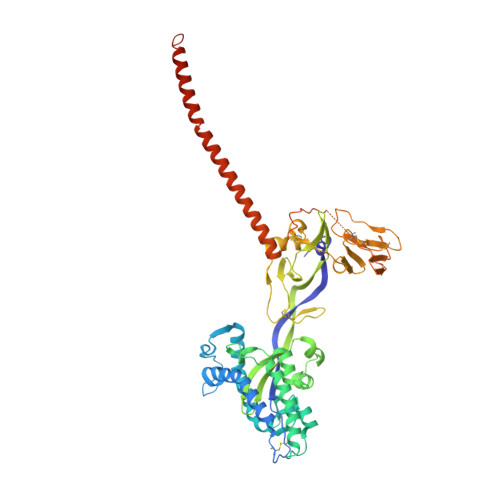
Structure of the parainfluenza virus 5 F protein in its metastable, prefusion conformation

Yin, H.-S.Wen, X.Paterson, R.G.Lamb, R.A.Jardetzky, T.S.

(2006) Nature 439: 38-44

  • DOI: https://doi.org/10.1038/nature04322
  • Primary Citation of Related Structures:  
    2B9B

  • PubMed Abstract: 

    Enveloped viruses have evolved complex glycoprotein machinery that drives the fusion of viral and cellular membranes, permitting entry of the viral genome into the cell. For the paramyxoviruses, the fusion (F) protein catalyses this membrane merger and entry step, and it has been postulated that the F protein undergoes complex refolding during this process. Here we report the crystal structure of the parainfluenza virus 5 F protein in its prefusion conformation, stabilized by the addition of a carboxy-terminal trimerization domain. The structure of the F protein shows that there are profound conformational differences between the pre- and postfusion states, involving transformations in secondary and tertiary structure. The positions and structural transitions of key parts of the fusion machinery, including the hydrophobic fusion peptide and two helical heptad repeat regions, clarify the mechanism of membrane fusion mediated by the F protein.


  • Organizational Affiliation

    Howard Hughes Medical Institute, Northwestern University, Evanston, Illinois 60208-3500, USA.


Macromolecules
Find similar proteins by:  (by identity cutoff)  |  3D Structure
Entity ID: 1
MoleculeChains Sequence LengthOrganismDetailsImage
Fusion glycoprotein F0
A, B, C
497Mammalian orthorubulavirus 5Mutation(s): 0 
Gene Names: F
UniProt
Find proteins for P04849 (Parainfluenza virus 5 (strain W3))
Explore P04849 
Go to UniProtKB:  P04849
Entity Groups  
Sequence Clusters30% Identity50% Identity70% Identity90% Identity95% Identity100% Identity
UniProt GroupP04849
Sequence Annotations
Expand
  • Reference Sequence
Small Molecules
Ligands 1 Unique
IDChains Name / Formula / InChI Key2D Diagram3D Interactions
NAG
Query on NAG

Download Ideal Coordinates CCD File 
D [auth A]
E [auth A]
F [auth A]
G [auth A]
H [auth B]
D [auth A],
E [auth A],
F [auth A],
G [auth A],
H [auth B],
I [auth B],
J [auth B],
K [auth B],
L [auth C],
M [auth C],
N [auth C],
O [auth C]
2-acetamido-2-deoxy-beta-D-glucopyranose
C8 H15 N O6
OVRNDRQMDRJTHS-FMDGEEDCSA-N
Experimental Data & Validation

Experimental Data

  • Method: X-RAY DIFFRACTION
  • Resolution: 2.85 Å
  • R-Value Free: 0.259 
  • R-Value Work: 0.222 
  • R-Value Observed: 0.222 
  • Space Group: C 2 2 21
Unit Cell:
Length ( Å )Angle ( ˚ )
a = 160.206α = 90
b = 259.864β = 90
c = 154.012γ = 90
Software Package:
Software NamePurpose
SCALAdata scaling
CNSrefinement
SHARPphasing
PDB_EXTRACTdata extraction
MOSFLMdata reduction
CCP4data scaling
SOLVEphasing
DMphasing
CNSphasing

Structure Validation

View Full Validation Report


Ligand Structure Quality Assessment 


Entry History 

Deposition Data

Revision History  (Full details and data files)

  • Version 1.0: 2006-01-24
    Type: Initial release
  • Version 1.1: 2008-05-01
    Changes: Version format compliance
  • Version 1.2: 2011-07-13
    Changes: Non-polymer description, Version format compliance
  • Version 1.3: 2020-07-29
    Type: Remediation
    Reason: Carbohydrate remediation
    Changes: Data collection, Database references, Derived calculations, Structure summary