High-resolution solution structure of the inhibitor-free catalytic fragment of human fibroblast collagenase determined by multidimensional NMR.
Moy, F.J., Chanda, P.K., Cosmi, S., Pisano, M.R., Urbano, C., Wilhelm, J., Powers, R.(1998) Biochemistry 37: 1495-1504
- PubMed: 9484219
- DOI: https://doi.org/10.1021/bi972181w
- Primary Citation of Related Structures:
1AYK, 2AYK - PubMed Abstract:
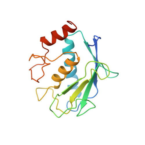
The high-resolution solution structure of the inhibitor-free catalytic fragment of human fibroblast collagenase (MMP-1), a protein of 18.7 kDa, which is a member of the matrix metalloproteinase family, has been determined using three-dimensional heteronuclear NMR spectroscopy. A total of 30 structures were calculated by means of hybrid distance geometry-simulated annealing using a total of 3333 experimental NMR restraints, consisting of 2409 approximate interproton distance restraints, 84 distance restraints for 42 backbone hydrogen bonds, 426 torsion angle restraints, 125 3JNH alpha restraints, 153 C alpha restraints, and 136 C beta restraints. The atomic rms distribution about the mean coordinate positions for the 30 structures for residues 7-137 and 145-163 is 0.42 +/- 0.04 A for the backbone atoms, 0.80 +/- 0.04 A for all atoms, and 0.50 +/- 0.03 A for all atoms excluding disordered side chains. The overall structure of MMP-1 is composed of a beta-sheet consisting of five beta-strands in a mixed parallel and anti-parallel arrangement and three alpha-helices. A best-fit superposition of the NMR structure of inhibitor-free MMP-1 with the 1.56 A resolution X-ray structure by Spurlino et al. [Spurlino, J. C., Smallwood, A. M., Carlton, D. D., Banks, T. M., Vavra, K. J., Johnson, J. S., Cook, E. R., Falvo, J., and Wahl, R. C., et al. (1994) Proteins: Struct., Funct., Genet. 19, 98-109] complexed with a hydroxamate inhibitor yields a backbone atomic rms difference of 1.22 A. The majority of differences between the NMR and X-ray structure occur in the vicinity of the active site for MMP-1. This includes an increase in mobility for residues 138-144 and a displacement for the Ca(2+)-loop (residues 74-80). Distinct differences were observed for side-chain torsion angles, in particular, the chi 1 for N80 is -60 degrees in the NMR structure compared to 180 degrees in the X-ray. This results in the side chain of N80 occupying and partially blocking access to the active site of MMP-1.
Organizational Affiliation:
Department of Structural Biology, Wyeth-Ayerst Research, Pearl River, New York 10965, USA.