2AVO

Kinetics, stability, and structural changes in high resolution crystal structures of HIV-1 protease with drug resistant mutations L24I, I50V, AND G73S


Experimental Data Snapshot

  • Method: X-RAY DIFFRACTION
  • Resolution: 1.10 Å
  • R-Value Free: 0.138 
  • R-Value Work: 0.108 
  • R-Value Observed: 0.107 

wwPDB Validation   3D Report Full Report


Ligand Structure Quality Assessment 


This is version 1.4 of the entry. See complete history


Literature

Kinetic, stability, and structural changes in high-resolution crystal structures of HIV-1 protease with drug-resistant mutations L24I, I50V, and G73S.

Liu, F.Boross, P.I.Wang, Y.F.Tozser, J.Louis, J.M.Harrison, R.W.Weber, I.T.

(2005) J Mol Biol 354: 789-800

  • DOI: https://doi.org/10.1016/j.jmb.2005.09.095
  • Primary Citation of Related Structures:  
    2AVM, 2AVO, 2AVQ, 2AVS, 2AVV

  • PubMed Abstract: 

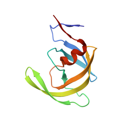
    The crystal structures, dimer stabilities, and kinetics have been analyzed for wild-type human immunodeficiency virus type 1 (HIV-1) protease (PR) and resistant mutants PR(L24I), PR(I50V), and PR(G73S) to gain insight into the molecular basis of drug resistance. The mutations lie in different structural regions. Mutation I50V alters a residue in the flexible flap that interacts with the inhibitor, L24I alters a residue adjacent to the catalytic Asp25, and G73S lies at the protein surface far from the inhibitor-binding site. PR(L24I) and PR(I50V), showed a 4% and 18% lower k(cat)/K(m), respectively, relative to PR. The relative k(cat)/K(m) of PR(G73S) varied from 14% to 400% when assayed using different substrates. Inhibition constants (K(i)) of the antiviral drug indinavir for the reaction catalyzed by the mutant enzymes were about threefold and 50-fold higher for PR(L24I) and PR(I50V), respectively, relative to PR and PR(G73S). The dimer dissociation constant (K(d)) was estimated to be approximately 20 nM for both PR(L24I) and PR(I50V), and below 5 nM for PR(G73S) and PR. Crystal structures of the mutants PR(L24I), PR(I50V) and PR(G73S) were determined in complexes with indinavir, or the p2/NC substrate analog at resolutions of 1.10-1.50 Angstrom. Each mutant revealed distinct structural changes relative to PR. The mutated residues in PR(L24I) and PR(I50V) had reduced intersubunit contacts, consistent with the increased K(d) for dimer dissociation. Relative to PR, PR(I50V) had fewer interactions of Val50 with inhibitors, in agreement with the dramatically increased K(i). The distal mutation G73S introduced new hydrogen bond interactions that can transmit changes to the substrate-binding site and alter catalytic activity. Therefore, the structural alterations observed for drug-resistant mutations were in agreement with kinetic and stability changes.


  • Organizational Affiliation

    Department of Biology, Molecular Basis of Disease Program, Georgia State University, Atlanta, GA 30303, USA.


Macromolecules
Find similar proteins by:  (by identity cutoff)  |  3D Structure
Entity ID: 1
MoleculeChains Sequence LengthOrganismDetailsImage
Pol polyprotein
A, B
99Human immunodeficiency virus 1Mutation(s): 6 
Gene Names: GAG-POL
EC: 3.4.23.16
UniProt
Find proteins for P04587 (Human immunodeficiency virus type 1 group M subtype B (isolate BH5))
Explore P04587 
Go to UniProtKB:  P04587
Find proteins for Q7SSI0 (Human immunodeficiency virus 1)
Explore Q7SSI0 
Go to UniProtKB:  Q7SSI0
Entity Groups  
Sequence Clusters30% Identity50% Identity70% Identity90% Identity95% Identity100% Identity
UniProt GroupQ7SSI0
Sequence Annotations
Expand
  • Reference Sequence
Small Molecules
Ligands 5 Unique
IDChains Name / Formula / InChI Key2D Diagram3D Interactions
MK1
Query on MK1

Download Ideal Coordinates CCD File 
E [auth B]N-[2(R)-HYDROXY-1(S)-INDANYL]-5-[(2(S)-TERTIARY BUTYLAMINOCARBONYL)-4(3-PYRIDYLMETHYL)PIPERAZINO]-4(S)-HYDROXY-2(R)-PHENYLMETHYLPENTANAMIDE
C36 H47 N5 O4
CBVCZFGXHXORBI-PXQQMZJSSA-N
SO4
Query on SO4

Download Ideal Coordinates CCD File 
C [auth A]SULFATE ION
O4 S
QAOWNCQODCNURD-UHFFFAOYSA-L
GOL
Query on GOL

Download Ideal Coordinates CCD File 
H [auth B]GLYCEROL
C3 H8 O3
PEDCQBHIVMGVHV-UHFFFAOYSA-N
DMS
Query on DMS

Download Ideal Coordinates CCD File 
D [auth A]DIMETHYL SULFOXIDE
C2 H6 O S
IAZDPXIOMUYVGZ-UHFFFAOYSA-N
ACY
Query on ACY

Download Ideal Coordinates CCD File 
F [auth B],
G [auth B]
ACETIC ACID
C2 H4 O2
QTBSBXVTEAMEQO-UHFFFAOYSA-N
Binding Affinity Annotations 
IDSourceBinding Affinity
MK1 BindingDB:  2AVO Ki: min: 0.05, max: 398 (nM) from 47 assay(s)
Kd: min: 1.07, max: 5.5 (nM) from 4 assay(s)
IC50: min: 0.36, max: 370 (nM) from 21 assay(s)
-TΔS: min: -8.28e+1, max: -2.80e+1 (kJ/mol) from 36 assay(s)
ΔH: min: -3.34e+1, max: 51 (kJ/mol) from 37 assay(s)
ΔG: min: -6.31e+1, max: -3.18e+1 (kJ/mol) from 35 assay(s)
PDBBind:  2AVO Ki: 1.4 (nM) from 1 assay(s)
Binding MOAD:  2AVO Ki: 1.4 (nM) from 1 assay(s)
Experimental Data & Validation

Experimental Data

  • Method: X-RAY DIFFRACTION
  • Resolution: 1.10 Å
  • R-Value Free: 0.138 
  • R-Value Work: 0.108 
  • R-Value Observed: 0.107 
  • Space Group: P 21 21 21
Unit Cell:
Length ( Å )Angle ( ˚ )
a = 51.465α = 90
b = 58.57β = 90
c = 61.644γ = 90
Software Package:
Software NamePurpose
ADSCdata collection
HKL-2000data reduction
AMoREphasing
SHELXL-97refinement
HKL-2000data scaling

Structure Validation

View Full Validation Report


Ligand Structure Quality Assessment 


Entry History 

Deposition Data

Revision History  (Full details and data files)

  • Version 1.0: 2006-01-24
    Type: Initial release
  • Version 1.1: 2008-05-01
    Changes: Version format compliance
  • Version 1.2: 2011-07-13
    Changes: Non-polymer description, Version format compliance
  • Version 1.3: 2021-10-20
    Changes: Database references, Derived calculations
  • Version 1.4: 2023-08-23
    Changes: Data collection, Refinement description