2AM4

Crystal Structure of N-Acetylglucosaminyltransferase I in Complex with UDP-2-deoxy-2-fluoro-glucose


Experimental Data Snapshot

  • Method: X-RAY DIFFRACTION
  • Resolution: 1.70 Å
  • R-Value Free: 0.217 
  • R-Value Work: 0.193 

wwPDB Validation   3D Report Full Report


Ligand Structure Quality Assessment 


This is version 1.3 of the entry. See complete history


Literature

X-ray Crystal Structures of Rabbit N-acetylglucosaminyltransferase I (GnT I) in Complex with Donor Substrate Analogues.

Gordon, R.D.Sivarajah, P.Satkunarajah, M.Ma, D.Tarling, C.A.Vizitiu, D.Withers, S.G.Rini, J.M.

(2006) J Mol Biol 360: 67-79

  • DOI: https://doi.org/10.1016/j.jmb.2006.04.058
  • Primary Citation of Related Structures:  
    2AM3, 2AM4, 2AM5, 2APC

  • PubMed Abstract: 

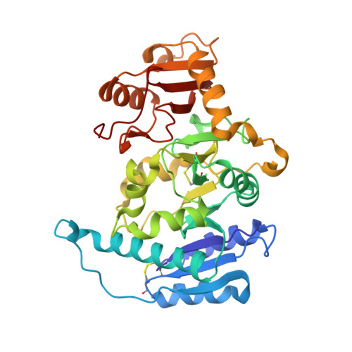
    The Golgi-resident glycosyltransferase, UDP-N-acetyl-d-glucosamine:alpha-3-d-mannoside beta-1,2-N-acetylglucosaminyltransferase I (GnT I), initiates the conversion of high-mannose oligosaccharides to complex and hybrid structures in the biosynthesis of N-linked glycans. Reported here are the X-ray crystal structures of GnT I in complex with UDP-CH2-GlcNAc (a non-hydrolyzable C-glycosidic phosphonate), UDP-2-deoxy-2-fluoro-glucose, UDP-glucose and UDP. Collectively, these structures provide evidence for the importance of the GlcNAc moiety and its N-acetyl group in donor substrate binding, as well as insight into the role played by the flexible 318-330 loop in substrate binding and product release. In addition, the UDP-CH2-GlcNAc complex reveals a well-defined glycerol molecule poised for nucleophilic attack on the C1 atom of the donor substrate analogue. The position and orientation of this glycerol molecule have allowed us to model the binding of the Manalpha1,3Manbeta1 moiety of the acceptor substrate and, based on the model, to suggest a rationalization for the main determinants of GnT I acceptor specificity.


  • Organizational Affiliation

    Department of Biochemistry, University of Toronto, Toronto, Ontario, Canada M5S 1A8.


Macromolecules
Find similar proteins by:  (by identity cutoff)  |  3D Structure
Entity ID: 1
MoleculeChains Sequence LengthOrganismDetailsImage
Alpha-1,3-mannosyl-glycoprotein 2-beta-N-acetylglucosaminyltransferase342Oryctolagus cuniculusMutation(s): 0 
Gene Names: MGAT1GNT1
EC: 2.4.1.101
UniProt
Find proteins for P27115 (Oryctolagus cuniculus)
Explore P27115 
Go to UniProtKB:  P27115
Entity Groups  
Sequence Clusters30% Identity50% Identity70% Identity90% Identity95% Identity100% Identity
UniProt GroupP27115
Sequence Annotations
Expand
  • Reference Sequence
Small Molecules
Binding Affinity Annotations 
IDSourceBinding Affinity
U2F PDBBind:  2AM4 Ki: 1.90e+5 (nM) from 1 assay(s)
Binding MOAD:  2AM4 Ki: 1.90e+5 (nM) from 1 assay(s)
Experimental Data & Validation

Experimental Data

  • Method: X-RAY DIFFRACTION
  • Resolution: 1.70 Å
  • R-Value Free: 0.217 
  • R-Value Work: 0.193 
  • Space Group: P 21 21 21
Unit Cell:
Length ( Å )Angle ( ˚ )
a = 40.917α = 90
b = 82.501β = 90
c = 102.394γ = 90
Software Package:
Software NamePurpose
CNSrefinement
DENZOdata reduction
SCALEPACKdata scaling
CNSphasing

Structure Validation

View Full Validation Report


Ligand Structure Quality Assessment 


Entry History 

Deposition Data

Revision History  (Full details and data files)

  • Version 1.0: 2006-06-27
    Type: Initial release
  • Version 1.1: 2008-04-30
    Changes: Version format compliance
  • Version 1.2: 2011-07-13
    Changes: Non-polymer description, Version format compliance
  • Version 1.3: 2023-08-23
    Changes: Data collection, Database references, Derived calculations, Refinement description