2ADA

ATOMIC STRUCTURE OF ADENOSINE DEAMINASE COMPLEXED WITH A TRANSITION-STATE ANALOG: UNDERSTANDING CATALYSIS AND IMMUNODEFICIENCY MUTATIONS


Experimental Data Snapshot

  • Method: X-RAY DIFFRACTION
  • Resolution: 2.40 Å
  • R-Value Work: 0.195 
  • R-Value Observed: 0.195 

wwPDB Validation   3D Report Full Report


Ligand Structure Quality Assessment 


This is version 1.3 of the entry. See complete history


Literature

Atomic structure of adenosine deaminase complexed with a transition-state analog: understanding catalysis and immunodeficiency mutations.

Wilson, D.K.Rudolph, F.B.Quiocho, F.A.

(1991) Science 252: 1278-1284

  • DOI: https://doi.org/10.1126/science.1925539
  • Primary Citation of Related Structures:  
    2ADA

  • PubMed Abstract: 

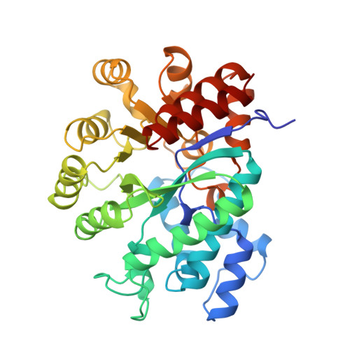
    The crystal structure of a murine adenosine deaminase complexed with 6-hydroxyl-1,6-dihydropurine ribonucleoside, a nearly ideal transition-state analog, has been determined and refined at 2.4 angstrom resolution. The structure is folded as an eight-stranded parallel alpha/beta barrel with a deep pocket at the beta-barrel COOH-terminal end wherein the inhibitor and a zinc are bound and completely sequestered. The presence of the zinc cofactor and the precise structure of the bound analog were not previously known. The 6R isomer of the analog is very tightly held in place by the coordination of the 6-hydroxyl to the zinc and the formation of nine hydrogen bonds. On the basis of the structure of the complex a stereoselective addition-elimination or SN2 mechanism of the enzyme is proposed with the zinc atom and the Glu and Asp residues playing key roles. A molecular explanation of a hereditary disease caused by several point mutations of an enzyme is also presented.


  • Organizational Affiliation

    Howard Hughes Medical Institute, Baylor College of Medicine, Houston, TX 77030.


Macromolecules
Find similar proteins by:  (by identity cutoff)  |  3D Structure
Entity ID: 1
MoleculeChains Sequence LengthOrganismDetailsImage
ADENOSINE DEAMINASE352Mus musculusMutation(s): 0 
EC: 3.5.4.4
UniProt
Find proteins for P03958 (Mus musculus)
Explore P03958 
Go to UniProtKB:  P03958
Entity Groups  
Sequence Clusters30% Identity50% Identity70% Identity90% Identity95% Identity100% Identity
UniProt GroupP03958
Sequence Annotations
Expand
  • Reference Sequence
Small Molecules
Ligands 2 Unique
IDChains Name / Formula / InChI Key2D Diagram3D Interactions
HPR
Query on HPR

Download Ideal Coordinates CCD File 
C [auth A]6-HYDROXY-7,8-DIHYDRO PURINE NUCLEOSIDE
C10 H14 N4 O5
ZWTNXGIZBOQCAJ-KQYNXXCUSA-N
ZN
Query on ZN

Download Ideal Coordinates CCD File 
B [auth A]ZINC ION
Zn
PTFCDOFLOPIGGS-UHFFFAOYSA-N
Binding Affinity Annotations 
IDSourceBinding Affinity
HPR Binding MOAD:  2ADA Ki: 1.00e-4 (nM) from 1 assay(s)
PDBBind:  2ADA Ki: 1.00e-4 (nM) from 1 assay(s)
Experimental Data & Validation

Experimental Data

  • Method: X-RAY DIFFRACTION
  • Resolution: 2.40 Å
  • R-Value Work: 0.195 
  • R-Value Observed: 0.195 
  • Space Group: C 1 2 1
Unit Cell:
Length ( Å )Angle ( ˚ )
a = 102.36α = 90
b = 94.11β = 127.19
c = 72.93γ = 90
Software Package:
Software NamePurpose
X-PLORmodel building
X-PLORrefinement
X-PLORphasing

Structure Validation

View Full Validation Report


Ligand Structure Quality Assessment 


Entry History 

Deposition Data

Revision History  (Full details and data files)

  • Version 1.0: 1995-03-31
    Type: Initial release
  • Version 1.1: 2008-03-24
    Changes: Version format compliance
  • Version 1.2: 2011-07-13
    Changes: Version format compliance
  • Version 1.3: 2024-02-14
    Changes: Data collection, Database references, Derived calculations, Other