2A9W

E. coli TS complexed with dUMP and inhibitor GA9


Experimental Data Snapshot

  • Method: X-RAY DIFFRACTION
  • Resolution: 1.65 Å
  • R-Value Free: 0.226 
  • R-Value Work: 0.207 
  • R-Value Observed: 0.207 

wwPDB Validation   3D Report Full Report


Ligand Structure Quality Assessment 


This is version 1.3 of the entry. See complete history


Literature

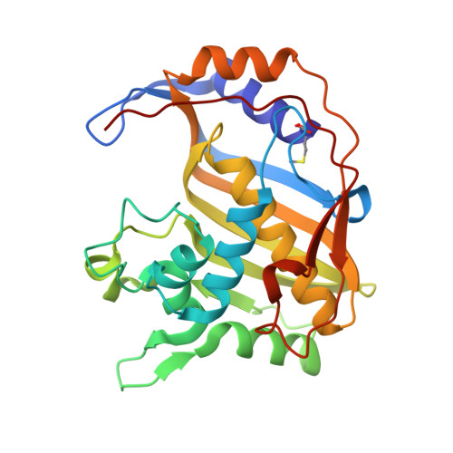
The structure of Cryptococcus neoformans thymidylate synthase suggests strategies for using target dynamics for species-specific inhibition.

Finer-Moore, J.S.Anderson, A.C.O'Neil, R.H.Costi, M.P.Ferrari, S.Krucinski, J.Stroud, R.M.

(2005) Acta Crystallogr D Biol Crystallogr 61: 1320-1334

  • DOI: https://doi.org/10.1107/S0907444905022638
  • Primary Citation of Related Structures:  
    2A9W, 2AAZ

  • PubMed Abstract: 

    The ternary complex crystal structures of Cryptococcus neoformans and Escherichia coli thymidylate synthase (TS) suggest mechanisms of species-specific inhibition of a highly conserved protein. The 2.1 Angstrom structure of C. neoformans TS cocrystallized with substrate and the cofactor analog CB3717 shows that the binding sites for substrate and cofactor are highly conserved with respect to human TS, but that the structure of the cofactor-binding site of C. neoformans TS is less constrained by surrounding residues. This feature might allow C. neoformans TS to form TS-dUMP-inhibitor complexes with a greater range of antifolates than human TS. 3',3''-Dibromophenol-4-chloro-1,8-naphthalein (GA9) selectively inhibits both E. coli TS and C. neoformans TS (K(i) = 4 microM) over human TS (K(i) >> 245 microM). The E. coli TS-dUMP-GA9 complex is in an open conformation, similar to that of the apoenzyme crystal structure. The GA9-binding site overlaps the binding site of the pABA-glutamyl moiety of the cofactor. The fact that human apoTS can adopt an unusual fold in which the GA9-binding site is disordered may explain the poor affinity of GA9 for the human enzyme. These observations highlight the critical need to incorporate multiple target conformations in any computational attempt to facilitate drug discovery.


  • Organizational Affiliation

    Department of Biochemistry and Biophysics, University of California, San Francisco, 600 16th Street, Room S412B, San Francisco, CA 94143-2240, USA.


Macromolecules
Find similar proteins by:  (by identity cutoff)  |  3D Structure
Entity ID: 1
MoleculeChains Sequence LengthOrganismDetailsImage
Thymidylate synthase
A, B, C, D
264Escherichia coliMutation(s): 1 
Gene Names: thyA
EC: 2.1.1.45
UniProt
Find proteins for P0A884 (Escherichia coli (strain K12))
Explore P0A884 
Go to UniProtKB:  P0A884
Entity Groups  
Sequence Clusters30% Identity50% Identity70% Identity90% Identity95% Identity100% Identity
UniProt GroupP0A884
Sequence Annotations
Expand
  • Reference Sequence
Small Molecules
Ligands 6 Unique
IDChains Name / Formula / InChI Key2D Diagram3D Interactions
GA9
Query on GA9

Download Ideal Coordinates CCD File 
BA [auth C],
H [auth A],
JA [auth D],
S [auth B]
3,3-BIS(3-BROMO-4-HYDROXYPHENYL)-7-CHLORO-1H,3H-BENZO[DE]ISOCHROMEN-1-ONE
C24 H13 Br2 Cl O4
GFGZCXHXQCQRFP-UHFFFAOYSA-N
UMP
Query on UMP

Download Ideal Coordinates CCD File 
AA [auth C],
G [auth A],
IA [auth D],
R [auth B]
2'-DEOXYURIDINE 5'-MONOPHOSPHATE
C9 H13 N2 O8 P
JSRLJPSBLDHEIO-SHYZEUOFSA-N
2BR
Query on 2BR

Download Ideal Coordinates CCD File 
T [auth B]2-BROMOPHENOL
C6 H5 Br O
VADKRMSMGWJZCF-UHFFFAOYSA-N
PO4
Query on PO4

Download Ideal Coordinates CCD File 
E [auth A]
F [auth A]
FA [auth D]
GA [auth D]
HA [auth D]
E [auth A],
F [auth A],
FA [auth D],
GA [auth D],
HA [auth D],
N [auth B],
O [auth B],
P [auth B],
Q [auth B],
X [auth C],
Y [auth C],
Z [auth C]
PHOSPHATE ION
O4 P
NBIIXXVUZAFLBC-UHFFFAOYSA-K
GOL
Query on GOL

Download Ideal Coordinates CCD File 
DA [auth C]
EA [auth C]
K [auth A]
L [auth A]
LA [auth D]
DA [auth C],
EA [auth C],
K [auth A],
L [auth A],
LA [auth D],
M [auth A],
MA [auth D],
U [auth B],
V [auth B],
W [auth B]
GLYCEROL
C3 H8 O3
PEDCQBHIVMGVHV-UHFFFAOYSA-N
BME
Query on BME

Download Ideal Coordinates CCD File 
CA [auth C],
I [auth A],
J [auth A],
KA [auth D]
BETA-MERCAPTOETHANOL
C2 H6 O S
DGVVWUTYPXICAM-UHFFFAOYSA-N
Modified Residues  1 Unique
IDChains TypeFormula2D DiagramParent
CXM
Query on CXM
A, B, C, D
L-PEPTIDE LINKINGC6 H11 N O4 SMET
Binding Affinity Annotations 
IDSourceBinding Affinity
GA9 Binding MOAD:  2A9W Ki: 4100 (nM) from 1 assay(s)
BindingDB:  2A9W Ki: 4100 (nM) from 1 assay(s)
Experimental Data & Validation

Experimental Data

  • Method: X-RAY DIFFRACTION
  • Resolution: 1.65 Å
  • R-Value Free: 0.226 
  • R-Value Work: 0.207 
  • R-Value Observed: 0.207 
  • Space Group: H 3
Unit Cell:
Length ( Å )Angle ( ˚ )
a = 186.6α = 90
b = 186.6β = 90
c = 114.2γ = 120
Software Package:
Software NamePurpose
CNSrefinement
HKL-2000data reduction
SCALEPACKdata scaling
AMoREphasing

Structure Validation

View Full Validation Report


Ligand Structure Quality Assessment 


Entry History 

Deposition Data

Revision History  (Full details and data files)

  • Version 1.0: 2005-10-11
    Type: Initial release
  • Version 1.1: 2008-04-30
    Changes: Version format compliance
  • Version 1.2: 2011-07-13
    Changes: Non-polymer description, Version format compliance
  • Version 1.3: 2023-08-23
    Changes: Data collection, Database references, Derived calculations, Refinement description