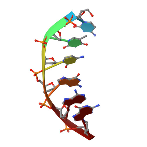
Crystal structure of the B-DNA hexamer d(CTCGAG): model for an A-to-B transition.
Wahl, M.C., Rao, S.T., Sundaralingam, M.(1996) Biophys J 70: 2857-2866
- PubMed: 8744323
- DOI: https://doi.org/10.1016/S0006-3495(96)79855-7
- Primary Citation of Related Structures:
251D - PubMed Abstract:
The crystal structure of the B-DNA hexamer d(CTCGAG) has been solved at 1.9 A resolution by iterative single isomorphous replacement, using the brominated derivative d(CG5BrCGAG), and refined to an R-factor of 18.6% for 120 nonhydrogen nucleic acid atoms and 32 water molecules. Although the central four base pairs form a typical B-form helix, several parameters suggest a transition to an A-like conformation at the termini. Based on this observation, a B-to-A transition was modeled, maintaining efficient base stacking across the junction. The wide minor groove (approximately 6.9 A) is reminiscent of that in the side-by-side double drug-DNA complexes and hosts a double spine of hydration. The global helix axes of the pseudo-continuous helices are at an acute angle of 60 degrees. The pseudocontinuous stacking is reinforced by the minor groove water structure extending between the two duplexes. The crossover point of two pairs of stacked duplexes is at the stacking junction, unlike that observed in the B-DNA decamers and dodecamers. This arrangement may have implications for the structure of a four-way DNA junction. The duplexes are arranged around a large (approximately 20 A diameter) channel centered on a 6(2) screw axis.
Organizational Affiliation:
Department of Chemistry, Ohio State University, Columbus 43210-1002, USA.