Crystal structures of four morpholino-doxorubicin anticancer drugs complexed with d(CGTACG) and d(CGATCG): implications in drug-DNA crosslink.
Gao, Y.G., Wang, A.H.(1995) J Biomol Struct Dyn 13: 103-117
- PubMed: 8527023
- DOI: https://doi.org/10.1080/07391102.1995.10508824
- Primary Citation of Related Structures:
215D, 234D, 235D, 236D - PubMed Abstract:
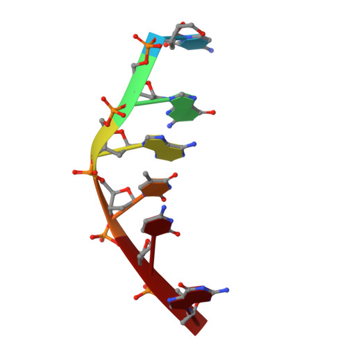
Among the new generations of anthracycline drugs, morpholino-doxorubicin (MDox) and its derivative have unusually potent activity when compared with the parent doxorubicin. 3"-Cyano-morpholino-doxorubicin (CN-MDox) has been suggested to form a covalent crosslink to DNA, although the exact mode of interactions remains unclear. To establish the structural basis of this crosslink, we carried out X-ray diffraction analyses of the complexes between four different morpholino-doxorubicins (i.e., MDox, CN-MDox, (R)- and (S)-2"-methoxy-morpholino-Dox (MMDox)) and two DNA hexamers CGTACG and CGATCG. Their crystal data are similar to other Dau/Dox complexes with space group P4(1)2(1)2,a = b approximately 28 A, c approximately 53 A. The refined structures at approximately 1.8 A resolution revealed that two drug molecules bind to the duplex with the aglycons intercalated between the CpG steps with their N3'-morpholino-daunosamines in the minor groove. The morpholino moiety is flexible and may adopt different conformations dependent on the sequence context. The O1" atoms of the two morpholino groups in the drug-DNA complexes are in van der Waals contact. The structural results suggest possible crosslinking mechanism of CN-MDox. It is worth pointing out that by linking two piperazinyl- or piperidinyl-doxorubicins at the 1" positions a new type of bis-doxorubicin derivatives may be synthesized which may bind to a hexanucleotide sequence with some specificity.
Organizational Affiliation:
Biophysics Division, University of Illinois at Urbana-Champaign 61801, USA.