Receptor-binding protein of Lactococcus lactis phages: identification and characterization of the saccharide receptor-binding site.
Tremblay, D.M., Tegoni, M., Spinelli, S., Campanacci, V., Blangy, S., Huyghe, C., Desmyter, A., Labrie, S., Moineau, S., Cambillau, C.(2006) J Bacteriol 188: 2400-2410
- PubMed: 16547026
- DOI: https://doi.org/10.1128/JB.188.7.2400-2410.2006
- Primary Citation of Related Structures:
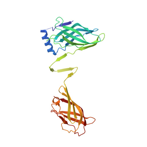
1ZRU - PubMed Abstract:
Phage p2, a member of the lactococcal 936 phage species, infects Lactococcus lactis strains by binding initially to specific carbohydrate receptors using its receptor-binding protein (RBP). The structures of p2 RBP, a homotrimeric protein composed of three domains, and of its complex with a neutralizing llama VH domain (VHH5) have been determined (S. Spinelli, A. Desmyter, C. T. Verrips, H. J. de Haard, S. Moineau, and C. Cambillau, Nat. Struct. Mol. Biol. 13:85-89, 2006). Here, we show that VHH5 was able to neutralize 12 of 50 lactococcal phages belonging to the 936 species. Moreover, escape phage mutants no longer neutralized by VHH5 were isolated from 11 of these phages. All of the mutations (but one) cluster in the RBP/VHH5 interaction surface that delineates the receptor-binding area. A glycerol molecule, observed in the 1.7-A resolution structure of RBP, was found to bind tightly (Kd= 0.26 microM) in a crevice located in this area. Other saccharides bind RBP with comparable high affinity. These data prove the saccharidic nature of the bacterial receptor recognized by phage p2 and identify the position of its binding site in the RBP head domain.
Organizational Affiliation:
Architecture et Fonction des Macromolécules Biologiques, UMR 6098 CNRS and Universités d'Aix-Marseille I & II, Campus de Luminy, case 932, 13288 Marseille CEDEX 09, France.