1Z3S

Angiopoietin-2 Receptor Binding Domain


Experimental Data Snapshot

  • Method: X-RAY DIFFRACTION
  • Resolution: 2.35 Å
  • R-Value Free: 0.287 
  • R-Value Work: 0.229 
  • R-Value Observed: 0.229 

wwPDB Validation   3D Report Full Report


This is version 1.2 of the entry. See complete history


Literature

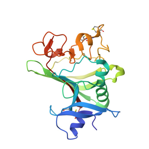
Structure of the angiopoietin-2 receptor binding domain and identification of surfaces involved in Tie2 recognition.

Barton, W.A.Tzvetkova, D.Nikolov, D.B.

(2005) Structure 13: 825-832

  • DOI: https://doi.org/10.1016/j.str.2005.03.009
  • Primary Citation of Related Structures:  
    1Z3S, 1Z3U

  • PubMed Abstract: 

    The angiopoietins comprise a small class of secreted glycoproteins that play crucial roles in the maturation and maintenance of the mammalian vascular and lymphatic systems. They exert their effects through a member of the tyrosine kinase receptor family, Tie2. Angiopoietin/Tie2 signaling is unique among tyrosine kinase receptor-ligand systems in that distinct angiopoietin ligands, although highly homologous, can function as agonists or antagonists in a context-dependent manner. In an effort to understand this molecular dichotomy, we have crystallized and determined the 2.4 A crystal structure of the Angiopoietin-2 (Ang2) receptor binding region. The structure reveals a fibrinogen fold with a unique C-terminal P domain. Conservation analysis and structure-based mutagenesis identify a groove on the Ang2 molecular surface that mediates receptor recognition.


  • Organizational Affiliation

    Structural Biology Program, Memorial Sloan-Kettering Cancer Center, 1275 York Avenue, New York, New York 10021, USA.


Macromolecules
Find similar proteins by:  (by identity cutoff)  |  3D Structure
Entity ID: 1
MoleculeChains Sequence LengthOrganismDetailsImage
Angiopoietin-2
A, B
217Homo sapiensMutation(s): 0 
UniProt & NIH Common Fund Data Resources
Find proteins for O15123 (Homo sapiens)
Explore O15123 
Go to UniProtKB:  O15123
PHAROS:  O15123
GTEx:  ENSG00000091879 
Entity Groups  
Sequence Clusters30% Identity50% Identity70% Identity90% Identity95% Identity100% Identity
UniProt GroupO15123
Sequence Annotations
Expand
  • Reference Sequence
Experimental Data & Validation

Experimental Data

  • Method: X-RAY DIFFRACTION
  • Resolution: 2.35 Å
  • R-Value Free: 0.287 
  • R-Value Work: 0.229 
  • R-Value Observed: 0.229 
  • Space Group: C 1 2 1
Unit Cell:
Length ( Å )Angle ( ˚ )
a = 74.19α = 90
b = 136.448β = 93.43
c = 47.732γ = 90
Software Package:
Software NamePurpose
DENZOdata reduction
SCALEPACKdata scaling
AMoREphasing
CNSrefinement

Structure Validation

View Full Validation Report


Entry History 

Deposition Data

Revision History  (Full details and data files)

  • Version 1.0: 2005-07-12
    Type: Initial release
  • Version 1.1: 2008-04-30
    Changes: Version format compliance
  • Version 1.2: 2011-07-13
    Changes: Version format compliance