1YP2

Crystal structure of potato tuber ADP-glucose pyrophosphorylase


Experimental Data Snapshot

  • Method: X-RAY DIFFRACTION
  • Resolution: 2.11 Å
  • R-Value Free: 0.229 
  • R-Value Work: 0.172 
  • R-Value Observed: 0.172 

wwPDB Validation   3D Report Full Report


Ligand Structure Quality Assessment 


This is version 1.2 of the entry. See complete history


Literature

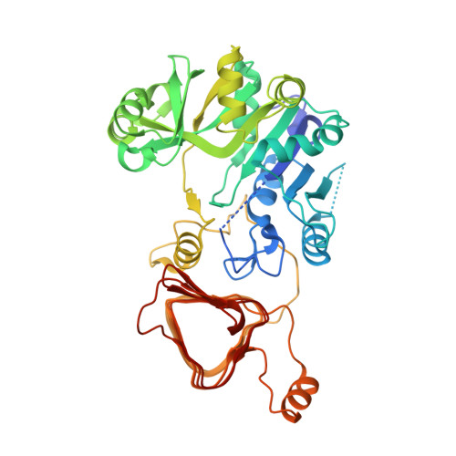
Crystal structure of potato tuber ADP-glucose pyrophosphorylase.

Jin, X.Ballicora, M.A.Preiss, J.Geiger, J.H.

(2005) EMBO J 24: 694-704

  • DOI: https://doi.org/10.1038/sj.emboj.7600551
  • Primary Citation of Related Structures:  
    1YP2, 1YP3, 1YP4

  • PubMed Abstract: 

    ADP-glucose pyrophosphorylase catalyzes the first committed and rate-limiting step in starch biosynthesis in plants and glycogen biosynthesis in bacteria. It is the enzymatic site for regulation of storage polysaccharide accumulation in plants and bacteria, being allosterically activated or inhibited by metabolites of energy flux. We report the first atomic resolution structure of ADP-glucose pyrophosphorylase. Crystals of potato tuber ADP-glucose pyrophosphorylase alpha subunit were grown in high concentrations of sulfate, resulting in the sulfate-bound, allosterically inhibited form of the enzyme. The N-terminal catalytic domain resembles a dinucleotide-binding Rossmann fold and the C-terminal domain adopts a left-handed parallel beta helix that is involved in cooperative allosteric regulation and a unique oligomerization. We also report structures of the enzyme in complex with ATP and ADP-glucose. Communication between the regulator-binding sites and the active site is both subtle and complex and involves several distinct regions of the enzyme including the N-terminus, the glucose-1-phosphate-binding site, and the ATP-binding site. These structures provide insights into the mechanism for catalysis and allosteric regulation of the enzyme.


  • Organizational Affiliation

    Department of Chemistry, Michigan State University, East Lansing, MI 48824, USA.


Macromolecules
Find similar proteins by:  (by identity cutoff)  |  3D Structure
Entity ID: 1
MoleculeChains Sequence LengthOrganismDetailsImage
Glucose-1-phosphate adenylyltransferase small subunit
A, B, C, D
451Solanum tuberosumMutation(s): 0 
EC: 2.7.7.27
UniProt
Find proteins for P23509 (Solanum tuberosum)
Explore P23509 
Go to UniProtKB:  P23509
Entity Groups  
Sequence Clusters30% Identity50% Identity70% Identity90% Identity95% Identity100% Identity
UniProt GroupP23509
Sequence Annotations
Expand
  • Reference Sequence
Small Molecules
Ligands 2 Unique
IDChains Name / Formula / InChI Key2D Diagram3D Interactions
PMB
Query on PMB

Download Ideal Coordinates CCD File 
H [auth A],
L [auth B],
P [auth C],
T [auth D]
PARA-MERCURY-BENZENESULFONIC ACID
C6 H5 Hg O3 S
KQAOIKIZSJJTII-UHFFFAOYSA-N
SO4
Query on SO4

Download Ideal Coordinates CCD File 
E [auth A]
F [auth A]
G [auth A]
I [auth B]
J [auth B]
E [auth A],
F [auth A],
G [auth A],
I [auth B],
J [auth B],
K [auth B],
M [auth C],
N [auth C],
O [auth C],
Q [auth D],
R [auth D],
S [auth D]
SULFATE ION
O4 S
QAOWNCQODCNURD-UHFFFAOYSA-L
Experimental Data & Validation

Experimental Data

  • Method: X-RAY DIFFRACTION
  • Resolution: 2.11 Å
  • R-Value Free: 0.229 
  • R-Value Work: 0.172 
  • R-Value Observed: 0.172 
  • Space Group: P 1 21 1
Unit Cell:
Length ( Å )Angle ( ˚ )
a = 79.465α = 90
b = 137.551β = 112.53
c = 91.623γ = 90
Software Package:
Software NamePurpose
CNSrefinement
HKL-2000data reduction
SCALEPACKdata scaling
SOLVEphasing

Structure Validation

View Full Validation Report


Ligand Structure Quality Assessment 


Entry History 

Deposition Data

Revision History  (Full details and data files)

  • Version 1.0: 2005-03-15
    Type: Initial release
  • Version 1.1: 2008-04-30
    Changes: Version format compliance
  • Version 1.2: 2011-07-13
    Changes: Version format compliance