1YLV

SCHIFF-BASE COMPLEX OF YEAST 5-AMINOLAEVULINIC ACID DEHYDRATASE WITH LAEVULINIC ACID


Experimental Data Snapshot

  • Method: X-RAY DIFFRACTION
  • Resolution: 2.15 Å
  • R-Value Free: 0.265 
  • R-Value Work: 0.207 

wwPDB Validation   3D Report Full Report


This is version 1.5 of the entry. See complete history


Literature

The Schiff base complex of yeast 5-aminolaevulinic acid dehydratase with laevulinic acid.

Erskine, P.T.Newbold, R.Roper, J.Coker, A.Warren, M.J.Shoolingin-Jordan, P.M.Wood, S.P.Cooper, J.B.

(1999) Protein Sci 8: 1250-1256

  • DOI: https://doi.org/10.1110/ps.8.6.1250
  • Primary Citation of Related Structures:  
    1YLV

  • PubMed Abstract: 

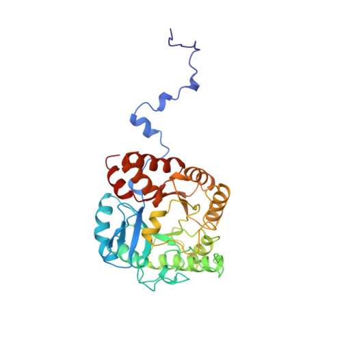
    The X-ray structure of the complex formed between yeast 5-aminolaevulinic acid dehydratase (ALAD) and the inhibitor laevulinic acid has been determined at 2.15 A resolution. The inhibitor binds by forming a Schiff base link with one of the two invariant lysines at the catalytic center: Lys263. It is known that this lysine forms a Schiff base link with substrate bound at the enzyme's so-called P-site. The carboxyl group of laevulinic acid makes hydrogen bonds with the side-chain-OH groups of Tyr329 and Ser290, as well as with the main-chain >NH group of Ser290. The aliphatic moiety of the inhibitor makes hydrophobic interactions with surrounding aromatic residues in the protein including Phe219, which resides in the flap covering the active site. Our analysis strongly suggests that the same interactions will be made by P-side substrate and also indicates that the substrate that binds at the enzyme's A-site will interact with the enzyme's zinc ion bound by three cysteines (133, 135, and 143). Inhibitor binding caused a substantial ordering of the active site flap (residues 217-235), which was largely invisible in the native electron density map and indicates that this highly conserved yet flexible region has a specific role in substrate binding during catalysis.


  • Organizational Affiliation

    Division of Biochemistry and Molecular Biology, School of Biological Sciences, University of Southampton, United Kingdom.


Macromolecules
Find similar proteins by:  (by identity cutoff)  |  3D Structure
Entity ID: 1
MoleculeChains Sequence LengthOrganismDetailsImage
PROTEIN (5-AMINOLAEVULINIC ACID DEHYDRATASE)342Saccharomyces cerevisiaeMutation(s): 0 
EC: 4.2.1.24
UniProt
Find proteins for P05373 (Saccharomyces cerevisiae (strain ATCC 204508 / S288c))
Explore P05373 
Go to UniProtKB:  P05373
Entity Groups  
Sequence Clusters30% Identity50% Identity70% Identity90% Identity95% Identity100% Identity
UniProt GroupP05373
Sequence Annotations
Expand
  • Reference Sequence
Small Molecules
Binding Affinity Annotations 
IDSourceBinding Affinity
SHF PDBBind:  1YLV Ki: 2.00e+6 (nM) from 1 assay(s)
Experimental Data & Validation

Experimental Data

  • Method: X-RAY DIFFRACTION
  • Resolution: 2.15 Å
  • R-Value Free: 0.265 
  • R-Value Work: 0.207 
  • Space Group: I 4 2 2
Unit Cell:
Length ( Å )Angle ( ˚ )
a = 103.4α = 90
b = 103.4β = 90
c = 168γ = 90
Software Package:
Software NamePurpose
CCP4model building
CCP4refinement
XDSdata reduction
CCP4data scaling
SCALAdata scaling
CCP4phasing

Structure Validation

View Full Validation Report


Entry History 

Deposition Data

Revision History  (Full details and data files)

  • Version 1.0: 2000-02-23
    Type: Initial release
  • Version 1.1: 2007-10-16
    Changes: Version format compliance
  • Version 1.2: 2011-07-13
    Changes: Derived calculations, Version format compliance
  • Version 1.3: 2018-04-04
    Changes: Data collection
  • Version 1.4: 2018-04-11
    Changes: Data collection
  • Version 1.5: 2023-08-23
    Changes: Data collection, Database references, Derived calculations, Refinement description