1Y2G

Crystal STructure of ZipA in complex with an inhibitor


Experimental Data Snapshot

  • Method: X-RAY DIFFRACTION
  • Resolution: 1.90 Å
  • R-Value Free: 0.241 
  • R-Value Work: 0.207 
  • R-Value Observed: 0.207 

wwPDB Validation   3D Report Full Report


This is version 1.3 of the entry. See complete history


Literature

A shape-based 3-D scaffold hopping method and its application to a bacterial protein-protein interaction

Rush, T.S.Grant, J.A.Mosyak, L.Nicholls, A.

(2005) J Med Chem 48: 1489-1495

  • DOI: https://doi.org/10.1021/jm040163o
  • Primary Citation of Related Structures:  
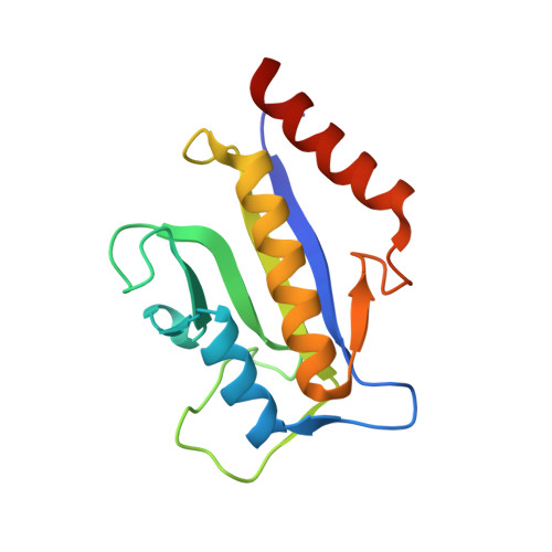
    1Y2F, 1Y2G

  • PubMed Abstract: 

    In this paper, we describe the first prospective application of the shape-comparison program ROCS (Rapid Overlay of Chemical Structures) to find new scaffolds for small molecule inhibitors of the ZipA-FtsZ protein-protein interaction, a proposed antibacterial target. The shape comparisons are made relative to the crystallographically determined, bioactive conformation of a high-throughput screening (HTS) hit. The use of ROCS led to the identification of a set of novel, weakly binding inhibitors with scaffolds presenting synthetic opportunities to further optimize biological affinity and lacking development issues associated with the HTS lead. These ROCS-identified scaffolds would have been missed using other structural similarity approaches such as ISIS 2D fingerprints. X-ray crystallographic analysis of one of the new inhibitors bound to ZipA reveals that the shape comparison approach very accurately predicted the binding mode. These experimental results validate this use of ROCS for chemotype switching or "lead hopping" and suggest that it is of general interest for lead identification in drug discovery endeavors.


  • Organizational Affiliation

    Department of Chemical & Screening Sciences, Wyeth Research, 87 Cambridge Park Drive, Cambridge, MA 02140, USA. trush@wyeth.com


Macromolecules
Find similar proteins by:  (by identity cutoff)  |  3D Structure
Entity ID: 1
MoleculeChains Sequence LengthOrganismDetailsImage
Cell division protein zipA
A, B
140Escherichia coliMutation(s): 0 
Gene Names: zipa
UniProt
Find proteins for P77173 (Escherichia coli (strain K12))
Explore P77173 
Go to UniProtKB:  P77173
Entity Groups  
Sequence Clusters30% Identity50% Identity70% Identity90% Identity95% Identity100% Identity
UniProt GroupP77173
Sequence Annotations
Expand
  • Reference Sequence
Small Molecules
Ligands 1 Unique
IDChains Name / Formula / InChI Key2D Diagram3D Interactions
CL3
Query on CL3

Download Ideal Coordinates CCD File 
C [auth A]N-METHYL-N-[3-(6-PHENYL[1,2,4]TRIAZOLO[4,3-B]PYRIDAZIN-3-YL)PHENYL]ACETAMIDE
C20 H17 N5 O
ALBWBHNFOJJMCV-UHFFFAOYSA-N
Binding Affinity Annotations 
IDSourceBinding Affinity
CL3 Binding MOAD:  1Y2G Kd: 8.31e+4 (nM) from 1 assay(s)
BindingDB:  1Y2G Kd: 8.31e+4 (nM) from 1 assay(s)
PDBBind:  1Y2G Kd: 8.31e+4 (nM) from 1 assay(s)
Experimental Data & Validation

Experimental Data

  • Method: X-RAY DIFFRACTION
  • Resolution: 1.90 Å
  • R-Value Free: 0.241 
  • R-Value Work: 0.207 
  • R-Value Observed: 0.207 
  • Space Group: P 1 21 1
Unit Cell:
Length ( Å )Angle ( ˚ )
a = 52.695α = 90
b = 39.521β = 104.97
c = 70.271γ = 90
Software Package:
Software NamePurpose
CNSrefinement

Structure Validation

View Full Validation Report


Entry History 

Deposition Data

Revision History  (Full details and data files)

  • Version 1.0: 2005-03-01
    Type: Initial release
  • Version 1.1: 2008-04-30
    Changes: Version format compliance
  • Version 1.2: 2011-07-13
    Changes: Version format compliance
  • Version 1.3: 2023-08-23
    Changes: Data collection, Database references, Derived calculations, Refinement description