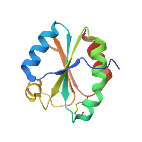
Comparative structural analysis of oxidized and reduced thioredoxin from Drosophila melanogaster
Wahl, M.C., Irmler, A., Hecker, B., Schirmer, R.H., Becker, K.(2005) J Mol Biol 345: 1119-1130
- PubMed: 15644209
- DOI: https://doi.org/10.1016/j.jmb.2004.11.004
- Primary Citation of Related Structures:
1XW9, 1XWA, 1XWB, 1XWC - PubMed Abstract:
Thioredoxins (Trx) participate in essential antioxidant and redox-regulatory processes via a pair of conserved cysteine residues. In dipteran insects like Drosophila and Anopheles, which lack a genuine glutathione reductase (GR), thioredoxins fuel the glutathione system with reducing equivalents. Thus, characterizing Trxs from these organisms contributes to our understanding of redox control in GR-free systems and provides information on novel targets for insect control. Cytosolic Trx of Drosophila melanogaster (DmTrx) is the first thioredoxin that was crystallized for X-ray diffraction analysis in the reduced and in the oxidized form. Comparison of the resulting structures shows rearrangements in the active-site regions. Formation of the C32-C35 disulfide bridge leads to a rotation of the side-chain of C32 away from C35 in the reduced form. This is similar to the situation in human Trx and Trx m from spinach chloroplasts but differs from Escherichia coli Trx, where it is C35 that moves upon change of the redox state. In all four crystal forms that were analysed, DmTrx molecules are engaged in a non-covalent dimer interaction. However, as demonstrated by gel-filtration analyses, DmTrx does not dimerize under quasi in vivo conditions and there is no redox control of a putative monomer/dimer equilibrium. The dimer dissociation constants K(d) were found to be 2.2mM for reduced DmTrx and above 10mM for oxidized DmTrx as well as for the protein in the presence of reduced glutathione. In human Trx, oxidative dimerization has been demonstrated in vitro. Therefore, this finding may indicate a difference in redox control of GR-free and GR-containing organisms.
Organizational Affiliation:
Max-Planck-Institut für Biophysikalische Chemie, Arbeitsgruppe Röntgenkristallographie, Am Fassberg 11, D-37077 Göttingen, Germany. mwahl@gwdg.de