1XC6

Native Structure Of Beta-Galactosidase from Penicillium sp. in complex with Galactose


Experimental Data Snapshot

  • Method: X-RAY DIFFRACTION
  • Resolution: 2.10 Å
  • R-Value Free: 0.186 
  • R-Value Work: 0.171 

wwPDB Validation   3D Report Full Report


Ligand Structure Quality Assessment 


This is version 2.0 of the entry. See complete history


Literature

Crystal Structures of beta-Galactosidase from Penicillium sp. and its Complex with Galactose

Rojas, A.L.Nagem, R.A.P.Neustroev, K.N.Arand, M.Adamska, M.Eneyskaya, E.V.Kulminskaya, A.A.Garratt, R.C.Golubev, A.M.Polikarpov, I.

(2004) J Mol Biol 343: 1281-1292

  • DOI: https://doi.org/10.1016/j.jmb.2004.09.012
  • Primary Citation of Related Structures:  
    1TG7, 1XC6

  • PubMed Abstract: 

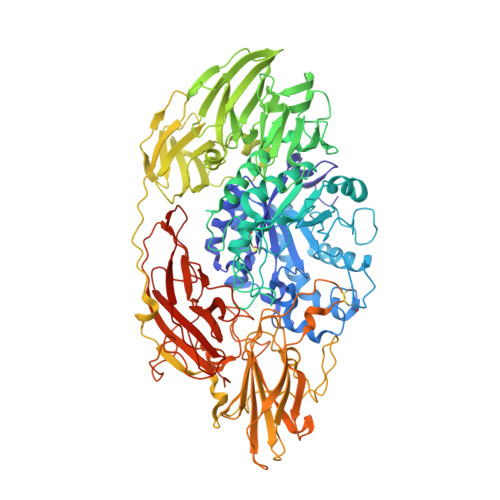
    Beta-galactosidases catalyze the hydrolysis of beta(1-3) and beta(1-4) galactosyl bonds in oligosaccharides as well as the inverse reaction of enzymatic condensation and transglycosylation. Here we report the crystallographic structures of Penicillium sp. beta-galactosidase and its complex with galactose solved by the SIRAS quick cryo-soaking technique at 1.90 A and 2.10 A resolution, respectively. The amino acid sequence of this 120 kDa protein was first assigned putatively on the basis of inspection of the experimental electron density maps and then determined by nucleotide sequence analysis. Primary structure alignments reveal that Penicillium sp. beta-galactosidase belongs to family 35 of glycosyl hydrolases (GHF-35). This model is the first 3D structure for a member of GHF-35. Five distinct domains which comprise the structure are assembled in a way previously unobserved for beta-galactosidases. Superposition of this complex with other beta-galactosidase complexes from several hydrolase families allowed the identification of residue Glu200 as the proton donor and residue Glu299 as the nucleophile involved in catalysis. Penicillium sp. beta-galactosidase is a glycoprotein containing seven N-linked oligosaccharide chains and is the only structure of a glycosylated beta-galactosidase described to date.


  • Organizational Affiliation

    Instituto de Física de São Carlos, Universidade de São Paulo, Av. Trabalhador São-carlense 400, CEP 13560-970 São Carlos, SP, Brazil.


Macromolecules
Find similar proteins by:  (by identity cutoff)  |  3D Structure
Entity ID: 1
MoleculeChains Sequence LengthOrganismDetailsImage
Beta-Galactosidase971Penicillium sp.Mutation(s): 0 
EC: 3.2.1.23
UniProt
Find proteins for Q700S9 (Penicillium sp)
Explore Q700S9 
Go to UniProtKB:  Q700S9
Entity Groups  
Sequence Clusters30% Identity50% Identity70% Identity90% Identity95% Identity100% Identity
UniProt GroupQ700S9
Sequence Annotations
Expand
  • Reference Sequence
Oligosaccharides

Help

Entity ID: 2
MoleculeChains Length2D Diagram Glycosylation3D Interactions
beta-D-mannopyranose-(1-4)-2-acetamido-2-deoxy-beta-D-glucopyranose-(1-4)-2-acetamido-2-deoxy-beta-D-glucopyranose
B
3N-Glycosylation
Glycosylation Resources
GlyTouCan:  G15407YE
GlyCosmos:  G15407YE
GlyGen:  G15407YE
Entity ID: 3
MoleculeChains Length2D Diagram Glycosylation3D Interactions
alpha-D-mannopyranose-(1-2)-alpha-D-mannopyranose-(1-3)-[alpha-D-mannopyranose-(1-2)-alpha-D-mannopyranose-(1-6)]alpha-D-mannopyranose-(1-6)-[beta-D-mannopyranose-(1-3)]beta-D-mannopyranose-(1-4)-2-acetamido-2-deoxy-beta-D-glucopyranose-(1-4)-2-acetamido-2-deoxy-beta-D-glucopyranose
C
9N-Glycosylation
Glycosylation Resources
GlyTouCan:  G72046TZ
GlyCosmos:  G72046TZ
GlyGen:  G72046TZ
Entity ID: 4
MoleculeChains Length2D Diagram Glycosylation3D Interactions
2-acetamido-2-deoxy-beta-D-glucopyranose-(1-4)-2-acetamido-2-deoxy-beta-D-glucopyranose
D
2N-Glycosylation
Glycosylation Resources
GlyTouCan:  G42666HT
GlyCosmos:  G42666HT
GlyGen:  G42666HT
Entity ID: 5
MoleculeChains Length2D Diagram Glycosylation3D Interactions
alpha-D-mannopyranose-(1-3)-alpha-D-mannopyranose-(1-6)-beta-D-mannopyranose-(1-4)-2-acetamido-2-deoxy-beta-D-glucopyranose-(1-4)-2-acetamido-2-deoxy-beta-D-glucopyranose
E
5N-Glycosylation
Glycosylation Resources
GlyTouCan:  G10756ZZ
GlyCosmos:  G10756ZZ
GlyGen:  G10756ZZ
Entity ID: 6
MoleculeChains Length2D Diagram Glycosylation3D Interactions
alpha-D-mannopyranose-(1-3)-[alpha-D-mannopyranose-(1-6)]alpha-D-mannopyranose-(1-6)-[alpha-D-mannopyranose-(1-3)]beta-D-mannopyranose-(1-4)-2-acetamido-2-deoxy-beta-D-glucopyranose-(1-4)-2-acetamido-2-deoxy-beta-D-glucopyranose
F
7N-Glycosylation
Glycosylation Resources
GlyTouCan:  G55220VL
GlyCosmos:  G55220VL
GlyGen:  G55220VL
Small Molecules
Ligands 6 Unique
IDChains Name / Formula / InChI Key2D Diagram3D Interactions
NAG
Query on NAG

Download Ideal Coordinates CCD File 
G [auth A],
H [auth A]
2-acetamido-2-deoxy-beta-D-glucopyranose
C8 H15 N O6
OVRNDRQMDRJTHS-FMDGEEDCSA-N
GAL
Query on GAL

Download Ideal Coordinates CCD File 
I [auth A]beta-D-galactopyranose
C6 H12 O6
WQZGKKKJIJFFOK-FPRJBGLDSA-N
IOD
Query on IOD

Download Ideal Coordinates CCD File 
AA [auth A]
AB [auth A]
BA [auth A]
BB [auth A]
CA [auth A]
AA [auth A],
AB [auth A],
BA [auth A],
BB [auth A],
CA [auth A],
CB [auth A],
DA [auth A],
DB [auth A],
EA [auth A],
EB [auth A],
FA [auth A],
FB [auth A],
GA [auth A],
GB [auth A],
HA [auth A],
HB [auth A],
IA [auth A],
IB [auth A],
JA [auth A],
JB [auth A],
KA [auth A],
KB [auth A],
L [auth A],
LA [auth A],
M [auth A],
MA [auth A],
N [auth A],
NA [auth A],
O [auth A],
OA [auth A],
P [auth A],
PA [auth A],
Q [auth A],
QA [auth A],
R [auth A],
RA [auth A],
S [auth A],
SA [auth A],
T [auth A],
TA [auth A],
U [auth A],
UA [auth A],
V [auth A],
VA [auth A],
W [auth A],
WA [auth A],
X [auth A],
XA [auth A],
Y [auth A],
YA [auth A],
Z [auth A],
ZA [auth A]
IODIDE ION
I
XMBWDFGMSWQBCA-UHFFFAOYSA-M
PO4
Query on PO4

Download Ideal Coordinates CCD File 
K [auth A]PHOSPHATE ION
O4 P
NBIIXXVUZAFLBC-UHFFFAOYSA-K
EDO
Query on EDO

Download Ideal Coordinates CCD File 
LB [auth A]
MB [auth A]
NB [auth A]
OB [auth A]
PB [auth A]
LB [auth A],
MB [auth A],
NB [auth A],
OB [auth A],
PB [auth A],
QB [auth A],
RB [auth A],
SB [auth A],
TB [auth A],
UB [auth A],
VB [auth A]
1,2-ETHANEDIOL
C2 H6 O2
LYCAIKOWRPUZTN-UHFFFAOYSA-N
NA
Query on NA

Download Ideal Coordinates CCD File 
J [auth A]SODIUM ION
Na
FKNQFGJONOIPTF-UHFFFAOYSA-N
Experimental Data & Validation

Experimental Data

  • Method: X-RAY DIFFRACTION
  • Resolution: 2.10 Å
  • R-Value Free: 0.186 
  • R-Value Work: 0.171 
  • Space Group: P 43
Unit Cell:
Length ( Å )Angle ( ˚ )
a = 110.96α = 90
b = 110.96β = 90
c = 161.05γ = 90
Software Package:
Software NamePurpose
DENZOdata reduction
SCALEPACKdata scaling
SHARPphasing
CNSrefinement

Structure Validation

View Full Validation Report


Ligand Structure Quality Assessment 


Entry History 

Revision History  (Full details and data files)

  • Version 1.0: 2004-11-02
    Type: Initial release
  • Version 1.1: 2008-04-30
    Changes: Version format compliance
  • Version 1.2: 2011-07-13
    Changes: Non-polymer description, Version format compliance
  • Version 1.3: 2017-12-20
    Changes: Advisory, Database references
  • Version 2.0: 2020-07-29
    Type: Remediation
    Reason: Carbohydrate remediation
    Changes: Atomic model, Data collection, Derived calculations, Structure summary