1X96

Crystal structure of Aldose Reductase with citrates bound in the active site


Experimental Data Snapshot

  • Method: X-RAY DIFFRACTION
  • Resolution: 1.40 Å
  • R-Value Free: 0.201 
  • R-Value Work: 0.161 
  • R-Value Observed: 0.161 

wwPDB Validation   3D Report Full Report


Ligand Structure Quality Assessment 


This is version 1.3 of the entry. See complete history


Literature

High-Resolution Structures of Human Aldose Reductase Holoenzyme in Complex with Stereoisomers of the Potent Inhibitor Fidarestat: Stereospecific Interaction between the Enzyme and a Cyclic Imide Type Inhibitor

El-Kabbani, O.Darmanin, C.Oka, M.Schulze-Briese, C.Tomizaki, T.Hazemann, I.Mitschler, A.Podjarny, A.

(2004) J Med Chem 47: 4530-4537

  • DOI: https://doi.org/10.1021/jm0497794
  • Primary Citation of Related Structures:  
    1X96, 1X97, 1X98

  • PubMed Abstract: 

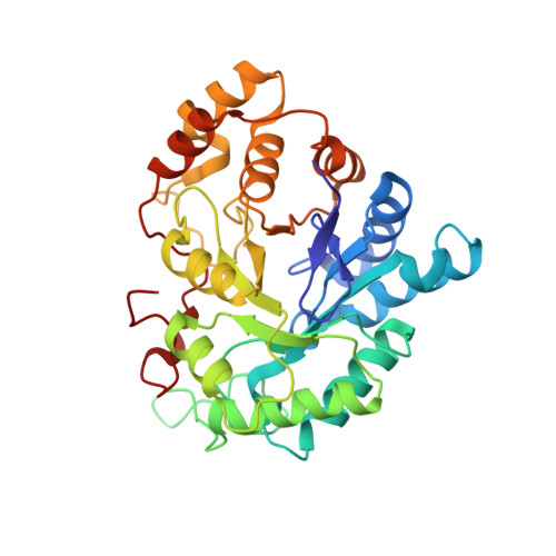
    Structure determinations of human aldose reductase holoenzyme in complex with the 2S4R-,2R4S- and 2R4R-isomers of the potent inhibitor Fidarestat ((2S,4S)-6-fluoro-2',5'-dioxospiro[chroman-4,4'-imidazoline]-2-carboxamide) were carried out in order to elucidate the binding modes responsible for the differences in their inhibitory potencies. In the complex structure with the 2R4S-isomer the cyclic imide moiety formed hydrogen bonds with the side-chains of Trp111, Tyr48 and His110. In the attempt to determine the complex structure with the least potent 2R4R-isomer this ligand was not observed, and instead, the active site was simultaneously occupied by two citrate molecules (occupancies of 60% and 40%). In the case of 2S4R, the active site was occupied by a citrate molecule which anchors the 2S4R-isomer from its carbamoyl group. The structures of the complexes suggest that the differences in the interactions between the cyclic imide rings and carbamoyl groups of the compounds with residues His110, Trp111, Trp219 and Cys298 account for differences in their inhibitory potencies.


  • Organizational Affiliation

    Department of Medicinal Chemistry, Victorian College of Pharmacy, Monash University (Parkville Campus), 381 Royal Parade, Parkville, Vic 3052, Australia. Ossama.El-Kabbani@vcp.monash.edu.au


Macromolecules
Find similar proteins by:  (by identity cutoff)  |  3D Structure
Entity ID: 1
MoleculeChains Sequence LengthOrganismDetailsImage
aldose reductase316Homo sapiensMutation(s): 0 
EC: 1.1.1.21
UniProt & NIH Common Fund Data Resources
Find proteins for P15121 (Homo sapiens)
Explore P15121 
Go to UniProtKB:  P15121
PHAROS:  P15121
GTEx:  ENSG00000085662 
Entity Groups  
Sequence Clusters30% Identity50% Identity70% Identity90% Identity95% Identity100% Identity
UniProt GroupP15121
Sequence Annotations
Expand
  • Reference Sequence
Experimental Data & Validation

Experimental Data

  • Method: X-RAY DIFFRACTION
  • Resolution: 1.40 Å
  • R-Value Free: 0.201 
  • R-Value Work: 0.161 
  • R-Value Observed: 0.161 
  • Space Group: P 1 21 1
Unit Cell:
Length ( Å )Angle ( ˚ )
a = 49.403α = 90
b = 66.782β = 92.03
c = 47.318γ = 90
Software Package:
Software NamePurpose
HKL-2000data collection
SCALEPACKdata scaling
X-PLORmodel building
SHELXL-97refinement
HKL-2000data reduction
X-PLORphasing

Structure Validation

View Full Validation Report


Ligand Structure Quality Assessment 


Entry History 

Deposition Data

Revision History  (Full details and data files)

  • Version 1.0: 2004-09-07
    Type: Initial release
  • Version 1.1: 2008-04-30
    Changes: Version format compliance
  • Version 1.2: 2011-07-13
    Changes: Version format compliance
  • Version 1.3: 2023-10-25
    Changes: Data collection, Database references, Derived calculations, Refinement description