1WX4

Crystal structure of the oxy-form of the copper-bound Streptomyces castaneoglobisporus tyrosinase complexed with a caddie protein prepared by the addition of dithiothreitol


Experimental Data Snapshot

  • Method: X-RAY DIFFRACTION
  • Resolution: 1.50 Å
  • R-Value Free: 0.216 
  • R-Value Work: 0.188 
  • R-Value Observed: 0.188 

wwPDB Validation   3D Report Full Report


This is version 1.4 of the entry. See complete history


Literature

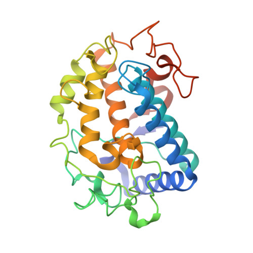
Crystallographic Evidence That the Dinuclear Copper Center of Tyrosinase Is Flexible during Catalysis

Matoba, Y.Kumagai, T.Yamamoto, A.Yoshitsu, H.Sugiyama, M.

(2006) J Biol Chem 281: 8981-8990

  • DOI: https://doi.org/10.1074/jbc.M509785200
  • Primary Citation of Related Structures:  
    1WX2, 1WX4, 1WX5, 1WXC, 2AHK, 2AHL, 2ZMX

  • PubMed Abstract: 

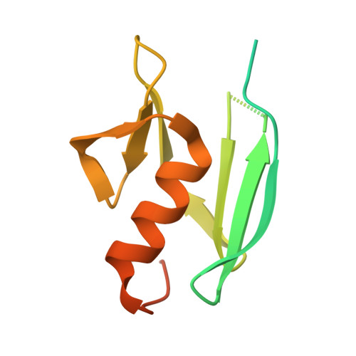
    At high resolution, we determined the crystal structures of copper-bound and metal-free tyrosinase in a complex with ORF378 designated as a "caddie" protein because it assists with transportation of two CuII ions into the tyrosinase catalytic center. These structures suggest that the caddie protein covers the hydrophobic molecular surface of tyrosinase and interferes with the binding of a substrate tyrosine to the catalytic site of tyrosinase. The caddie protein, which consists of one six-strandedbeta-sheet and one alpha-helix, has no similarity with all proteins deposited into the Protein Data Bank. Although tyrosinase and catechol oxidase are classified into the type 3 copper protein family, the latter enzyme lacks monooxygenase activity. The difference in catalytic activity is based on the structural observations that a large vacant space is present just above the active center of tyrosinase and that one of the six His ligands for the two copper ions is highly flexible. These structural characteristics of tyrosinase suggest that, in the reaction that catalyzes the ortho-hydroxylation of monophenol, one of the two Cu(II) ions is coordinated by the peroxide-originated oxygen bound to the substrate. Our crystallographic study shows evidence that the tyrosinase active center formed by dinuclear coppers is flexible during catalysis.


  • Organizational Affiliation

    Department of Molecular Microbiology and Biotechnology, Graduate School of Biomedical Sciences, Hiroshima University, Hiroshima, Japan.


Macromolecules
Find similar proteins by:  (by identity cutoff)  |  3D Structure
Entity ID: 1
MoleculeChains Sequence LengthOrganismDetailsImage
tyrosinase281Streptomyces castaneoglobisporusMutation(s): 0 
Gene Names: tyrc
EC: 1.14.18.1
UniProt
Find proteins for Q83WS2 (Streptomyces castaneoglobisporus)
Explore Q83WS2 
Go to UniProtKB:  Q83WS2
Entity Groups  
Sequence Clusters30% Identity50% Identity70% Identity90% Identity95% Identity100% Identity
UniProt GroupQ83WS2
Sequence Annotations
Expand
  • Reference Sequence
Find similar proteins by:  (by identity cutoff)  |  3D Structure
Entity ID: 2
MoleculeChains Sequence LengthOrganismDetailsImage
MelC134Streptomyces castaneoglobisporusMutation(s): 0 
Gene Names: orf378
UniProt
Find proteins for Q83WS1 (Streptomyces castaneoglobisporus)
Explore Q83WS1 
Go to UniProtKB:  Q83WS1
Entity Groups  
Sequence Clusters30% Identity50% Identity70% Identity90% Identity95% Identity100% Identity
UniProt GroupQ83WS1
Sequence Annotations
Expand
  • Reference Sequence
Small Molecules
Ligands 3 Unique
IDChains Name / Formula / InChI Key2D Diagram3D Interactions
CU
Query on CU

Download Ideal Coordinates CCD File 
C [auth A],
D [auth A],
E [auth A],
J [auth B]
COPPER (II) ION
Cu
JPVYNHNXODAKFH-UHFFFAOYSA-N
NO3
Query on NO3

Download Ideal Coordinates CCD File 
F [auth A],
G [auth A],
H [auth A],
K [auth B],
L [auth B]
NITRATE ION
N O3
NHNBFGGVMKEFGY-UHFFFAOYSA-N
PER
Query on PER

Download Ideal Coordinates CCD File 
I [auth A]PEROXIDE ION
O2
ANAIPYUSIMHBEL-UHFFFAOYSA-N
Experimental Data & Validation

Experimental Data

  • Method: X-RAY DIFFRACTION
  • Resolution: 1.50 Å
  • R-Value Free: 0.216 
  • R-Value Work: 0.188 
  • R-Value Observed: 0.188 
  • Space Group: P 21 21 2
Unit Cell:
Length ( Å )Angle ( ˚ )
a = 65.36α = 90
b = 98.23β = 90
c = 55.17γ = 90
Software Package:
Software NamePurpose
X-PLORrefinement
MOSFLMdata reduction
CCP4data scaling
X-PLORphasing

Structure Validation

View Full Validation Report


Entry History 

Deposition Data

Revision History  (Full details and data files)

  • Version 1.0: 2006-01-31
    Type: Initial release
  • Version 1.1: 2008-04-30
    Changes: Version format compliance
  • Version 1.2: 2011-07-13
    Changes: Version format compliance
  • Version 1.3: 2011-11-16
    Changes: Atomic model
  • Version 1.4: 2023-10-25
    Changes: Data collection, Database references, Derived calculations, Refinement description