1WNG

Structural study of project ID PH0725 from Pyrococcus horikoshii OT3


Experimental Data Snapshot

  • Method: X-RAY DIFFRACTION
  • Resolution: 2.10 Å
  • R-Value Free: 0.228 
  • R-Value Work: 0.198 
  • R-Value Observed: 0.198 

wwPDB Validation   3D Report Full Report


Ligand Structure Quality Assessment 


This is version 1.3 of the entry. See complete history


Literature

Structures of two archaeal diphthine synthases: insights into the post-translational modification of elongation factor 2.

Kishishita, S.Shimizu, K.Murayama, K.Terada, T.Shirouzu, M.Yokoyama, S.Kunishima, N.

(2008) Acta Crystallogr D Biol Crystallogr 64: 397-406

  • DOI: https://doi.org/10.1107/S0907444908000723
  • Primary Citation of Related Structures:  
    1WDE, 1WNG

  • PubMed Abstract: 

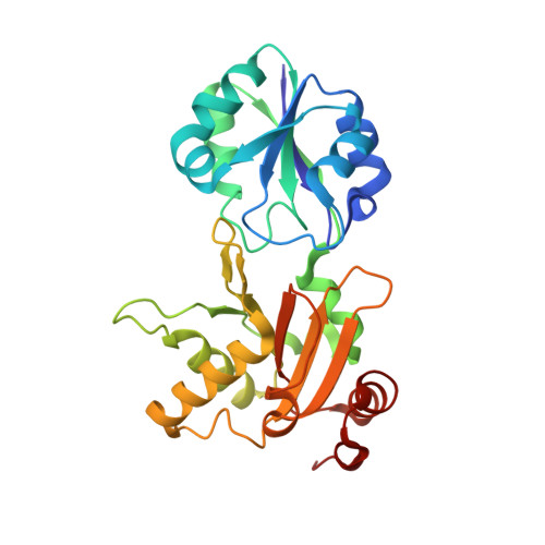
    The target of diphtheria toxin is the diphthamide residue in translation elongation factor 2 (EF-2), which is generated by a three-step post-translational modification of a specific histidine residue in the EF-2 precursor. In the second modification step, an S-adenosylmethionine-dependent methyltransferase, diphthine synthase (DS), catalyzes the trimethylation of the EF-2 precursor. The homodimeric crystal structures of the archaeal diphthine synthases from Pyrococcus horikoshii OT3 and Aeropyrum pernix K1 have been determined. These structures share essentially the same overall fold as the cobalt-precorrin-4 methyltransferase CbiF, confirming that DS belongs to the dimeric class III family of methyltransferases. In the P. horikoshii DS dimer, only one of the two active sites binds the reaction product S-adenosyl-L-homocysteine (AdoHcy), while the other active site contains no ligand. This asymmetric AdoHcy binding may be a consequence of intra-domain and inter-domain movements upon binding of AdoHcy at one of the two sites. These movements disrupt the twofold dimeric symmetry of the DS dimer and probably cause lower AdoHcy affinity at the other binding site.


  • Organizational Affiliation

    Protein Research Group, Genomic Sciences Center, Yokohama Institute, RIKEN 1-7-22, Suehiro-cho, Tsurumi, Yokohama 230-0045, Japan.


Macromolecules
Find similar proteins by:  (by identity cutoff)  |  3D Structure
Entity ID: 1
MoleculeChains Sequence LengthOrganismDetailsImage
Probable diphthine synthase
A, B
265Pyrococcus horikoshiiMutation(s): 0 
EC: 2.1.1.98
UniProt
Find proteins for O58456 (Pyrococcus horikoshii (strain ATCC 700860 / DSM 12428 / JCM 9974 / NBRC 100139 / OT-3))
Explore O58456 
Go to UniProtKB:  O58456
Entity Groups  
Sequence Clusters30% Identity50% Identity70% Identity90% Identity95% Identity100% Identity
UniProt GroupO58456
Sequence Annotations
Expand
  • Reference Sequence
Experimental Data & Validation

Experimental Data

  • Method: X-RAY DIFFRACTION
  • Resolution: 2.10 Å
  • R-Value Free: 0.228 
  • R-Value Work: 0.198 
  • R-Value Observed: 0.198 
  • Space Group: P 41 21 2
Unit Cell:
Length ( Å )Angle ( ˚ )
a = 104.914α = 90
b = 104.914β = 90
c = 137.34γ = 90
Software Package:
Software NamePurpose
CNSrefinement
HKL-2000data reduction
SCALEPACKdata scaling
MOLREPphasing

Structure Validation

View Full Validation Report


Ligand Structure Quality Assessment 


Entry History 

Deposition Data

Revision History  (Full details and data files)

  • Version 1.0: 2005-07-19
    Type: Initial release
  • Version 1.1: 2008-04-30
    Changes: Version format compliance
  • Version 1.2: 2011-07-13
    Changes: Version format compliance
  • Version 1.3: 2024-03-13
    Changes: Data collection, Database references, Derived calculations