1W45

The 2.5 Angstroem structure of the K16A mutant of annexin A8, which has an intact N-terminus.


Experimental Data Snapshot

  • Method: X-RAY DIFFRACTION
  • Resolution: 2.51 Å
  • R-Value Free: 0.269 
  • R-Value Work: 0.206 
  • R-Value Observed: 0.209 

wwPDB Validation   3D Report Full Report


This is version 1.4 of the entry. See complete history


Literature

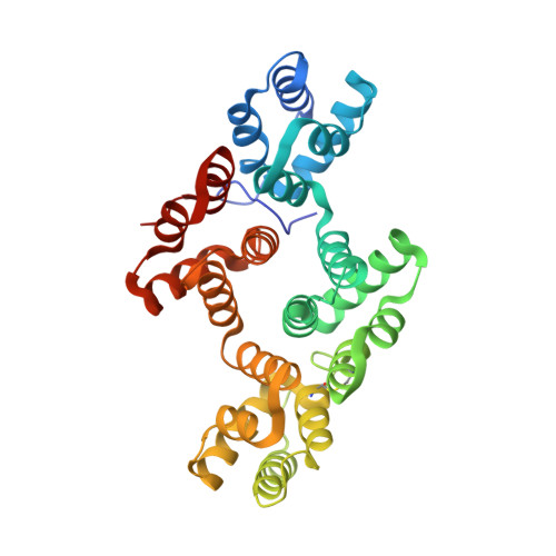
The Crystal Structure of Annexin A8 is Similar to that of Annexin A3

Rety, S.Sopkova-De Oliveira Santos, J.Dreyfuss, L.Blondeau, K.Hofbauerova, K.Raguenes-Nicol, C.Kerboeuf, D.Renouard, M.Russo-Marie, F.Lewit-Bentley, A.

(2005) J Mol Biol 345: 1131

  • DOI: https://doi.org/10.1016/j.jmb.2004.11.015
  • Primary Citation of Related Structures:  
    1W3W, 1W45

  • PubMed Abstract: 

    Annexin A8 is a relatively infrequent and poorly studied member of this large family of calcium-binding and membrane-binding proteins. It is, however, associated with a specific disease, acute promyelocytic leukemia. We have solved its three-dimensional structure, which includes a moderately long and intact N terminus. The structure is closest to that of annexin A3 and highlights several important regions of inherent flexibility in the annexin molecule. The N terminus resembles that of annexin A3, as it lies along the concave surface of the molecule and inserts partially into the hydrophilic channel in its centre. Since both annexins A3 and A8 are expressed in promyelocytic cells during their differentiation, the similarity in their structures might suggest a functional relationship.


  • Organizational Affiliation

    LURE, Centre Universitaire Paris-Sud, BP 34, 91898 Orsay Cedex, France.


Macromolecules
Find similar proteins by:  (by identity cutoff)  |  3D Structure
Entity ID: 1
MoleculeChains Sequence LengthOrganismDetailsImage
ANNEXIN A8
A, B
327Homo sapiensMutation(s): 1 
UniProt & NIH Common Fund Data Resources
Find proteins for P13928 (Homo sapiens)
Explore P13928 
Go to UniProtKB:  P13928
PHAROS:  P13928
GTEx:  ENSG00000265190 
Entity Groups  
Sequence Clusters30% Identity50% Identity70% Identity90% Identity95% Identity100% Identity
UniProt GroupP13928
Sequence Annotations
Expand
  • Reference Sequence
Experimental Data & Validation

Experimental Data

  • Method: X-RAY DIFFRACTION
  • Resolution: 2.51 Å
  • R-Value Free: 0.269 
  • R-Value Work: 0.206 
  • R-Value Observed: 0.209 
  • Space Group: P 1
Unit Cell:
Length ( Å )Angle ( ˚ )
a = 49.297α = 84.42
b = 59.653β = 83.32
c = 70.788γ = 74.1
Software Package:
Software NamePurpose
REFMACrefinement
DENZOdata reduction
SCALEPACKdata scaling
AMoREphasing

Structure Validation

View Full Validation Report


Entry History 

Revision History  (Full details and data files)

  • Version 1.0: 2005-01-18
    Type: Initial release
  • Version 1.1: 2011-05-08
    Changes: Version format compliance
  • Version 1.2: 2011-07-13
    Changes: Version format compliance
  • Version 1.3: 2017-06-28
    Changes: Data collection
  • Version 1.4: 2023-12-13
    Changes: Data collection, Database references, Other, Refinement description