


Structure of oxygen-bound form of a Heme PAS domain of Ec DOS
Kurokawa, H., Watanabe, M., Sagami, I., Mikami, B., Shimizu, T.To be published.
Experimental Data Snapshot
Entity ID: 1 | |||||
|---|---|---|---|---|---|
| Molecule | Chains | Sequence Length | Organism | Details | Image |
| Heme pas sensor protein | 167 | Escherichia coli K-12 | Mutation(s): 0 | 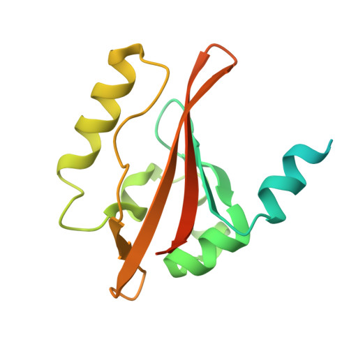 | |
UniProt | |||||
Find proteins for P76129 (Escherichia coli (strain K12)) Explore P76129 Go to UniProtKB: P76129 | |||||
Entity Groups | |||||
| Sequence Clusters | 30% Identity50% Identity70% Identity90% Identity95% Identity100% Identity | ||||
| UniProt Group | P76129 | ||||
Sequence AnnotationsExpand | |||||
| |||||
| Ligands 2 Unique | |||||
|---|---|---|---|---|---|
| ID | Chains | Name / Formula / InChI Key | 2D Diagram | 3D Interactions | |
| HEM Query on HEM | C [auth A], D [auth B] | PROTOPORPHYRIN IX CONTAINING FE C34 H32 Fe N4 O4 KABFMIBPWCXCRK-RGGAHWMASA-L |  | ||
| OXY Query on OXY | E [auth B] | OXYGEN MOLECULE O2 MYMOFIZGZYHOMD-UHFFFAOYSA-N |  | ||
| Length ( Å ) | Angle ( ˚ ) |
|---|---|
| a = 47.894 | α = 90 |
| b = 69.337 | β = 90 |
| c = 81.876 | γ = 90 |
| Software Name | Purpose |
|---|---|
| CNS | refinement |
| DENZO | data reduction |
| SCALEPACK | data scaling |
| CNS | phasing |
RCSB PDB (citation) is hosted by
RCSB PDB is a member of the

