Structural aspects of interfacial adsorption. A crystallographic and site-directed mutagenesis study of the phospholipase A2 from the venom of Agkistrodon piscivorus piscivorus.
Han, S.K., Yoon, E.T., Scott, D.L., Sigler, P.B., Cho, W.(1997) J Biol Chem 272: 3573-3582
- PubMed: 9013608
- Primary Citation of Related Structures:
1VAP - PubMed Abstract:
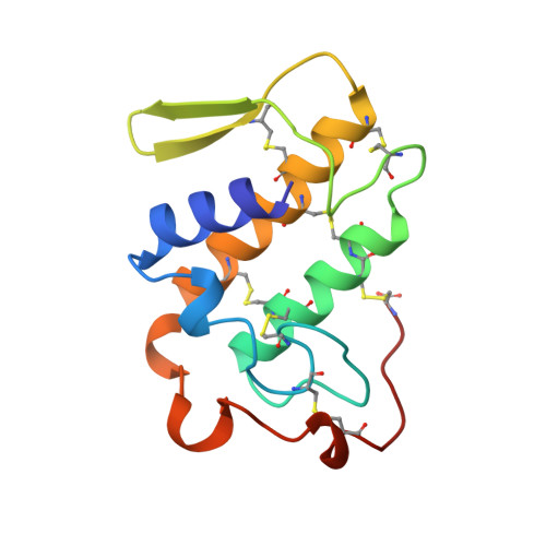
Recent genetic and structural studies have shed considerable light on the mechanism by which secretory phospholipases A2 interact with substrate aggregates. Electrostatic forces play an essential role in optimizing interfacial catalysis. Efficient and productive adsorption of the Class I bovine pancreatic phospholipase A2 to anionic interfaces is dependent upon the presence of two nonconserved lysine residues at sequence positions 56 and 116, implying that critical components of the adsorption surface differ among enzyme species (Dua, R., Wu, S.-K., and Cho, W. (1995) J. Biol. Chem. 270, 263-268). In an effort to further characterize the protein residues involved in interfacial catalysis, we have determined the high resolution (1.7 A) x-ray structure of the Class II Asp-49 phospholipase A2 from the venom of Agkistrodon piscivorus piscivorus. Correlation of the three-dimensional coordinates with kinetic data derived from site-directed mutations near the amino terminus (E6R, K7E, K10E, K11E, and K16E) and the active site (K54E and K69Y) defines much of the interface topography. Lysine residues at sequence positions 7 and 10 mediate the adsorption of A. p. piscivorus phospholipase A2 to anionic interfaces but play little role in the enzyme's interaction with electrically neutral surfaces or in substrate binding. Compared to the native enzyme, the mutant proteins K7E and K10E demonstrate comparable (20-fold) decreases in affinity and catalysis on polymerized mixed liposomes of 1-hexadecanoyl-2-(1-pyrenedecanoyl)-sn-glycero-3-phosphocholine and 1,2-bis[12-(lipoyloxy)dodecanoyl]-sn-glycero-3-phosphoglycerol, while the double mutant, K7E/K10E, shows a more dramatic 500-fold decrease in catalysis and interfacial adsorption. The calculated contributions of Lys-7 and Lys-10 to the free energy of binding of A. p. piscivorus phospholipase A2 to anionic liposomes (-1.8 kcal/mol at 25 degrees C per lysine) are additive (i.e. -3.7 kcal/mol) and together represent nearly half of the total binding energy. Although both lysine side chains lie exposed at the edge of the proposed interfacial adsorption surface, they are geographically remote from the corresponding interfacial determinants for the bovine enzyme. Our results confirm that interfacial adsorption is largely driven by electrostatic forces and demonstrate that the arrangement of the critical charges (e.g. lysines) is species-specific. This variability in the topography of the adsorption surface suggests a corresponding flexibility in the orientation of the active enzyme at the substrate interface.
Organizational Affiliation:
Department of Chemistry, University of Illinois at Chicago, Chicago, Illinois 60607-7061, USA.