1V9A

Crystal structure of Uroporphyrin-III C-methyl transferase from Thermus thermophilus complexed with S-adenyl homocysteine


Experimental Data Snapshot

  • Method: X-RAY DIFFRACTION
  • Resolution: 2.00 Å
  • R-Value Free: 0.246 
  • R-Value Work: 0.215 
  • R-Value Observed: 0.215 

wwPDB Validation   3D Report Full Report


Ligand Structure Quality Assessment 


This is version 1.3 of the entry. See complete history


Literature

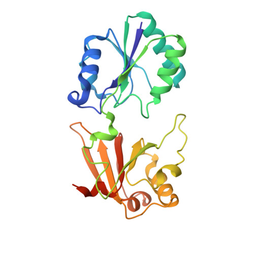
Structure of a closed-form uroporphyrinogen-III C-methyltransferase from Thermus thermophilus.

Rehse, P.H.Kitao, T.Tahirov, T.H.

(2005) Acta Crystallogr D Biol Crystallogr 61: 913-919

  • DOI: https://doi.org/10.1107/S0907444905008838
  • Primary Citation of Related Structures:  
    1V9A, 1VA0

  • PubMed Abstract: 

    Uroporphyrinogen-III C-methyltransferase from Thermus thermophilus is a multifunctional protein responsible for two of the eight S-adenosyl-methionine-dependent methylations of the corrin ring during vitamin B(12) synthesis. The structure of this protein has been solved to 2.0 A resolution in both the apo and cofactor-bound form. The monomer consists of two domains, A and B, each consisting of a five-stranded beta-sheet and two or three alpha-helices, with the cofactor bound at the interface. The biological unit is the dimer found in the asymmetric unit. This dimer is related by a non-crystallographic twofold such that two B domains combine to form a long ten-stranded beta-sheet. When compared with solved related structures, this structure shows clear differences in the region involved in cofactor and substrate binding, affirming the role of several previously implicated residues and questioning others. The solved related structures are characterized by an exposed active site. The T. thermophilus structure has this site restricted by the interaction of a flexible loop structure with a highly conserved residue, suggesting a mechanistic role. This structure represents the ;closed' form of the protein.


  • Organizational Affiliation

    APCG, RIKEN Harima Institute, 1-1-1 Kouto, Mikazuki-cho, Sayo-gun, Hyogo 679-5148, Japan.


Macromolecules
Find similar proteins by:  (by identity cutoff)  |  3D Structure
Entity ID: 1
MoleculeChains Sequence LengthOrganismDetailsImage
Uroporphyrin-III C-methyltransferase
A, B
239Thermus thermophilus HB8Mutation(s): 0 
EC: 2.1.1.107
UniProt
Find proteins for Q5SKH6 (Thermus thermophilus (strain ATCC 27634 / DSM 579 / HB8))
Explore Q5SKH6 
Go to UniProtKB:  Q5SKH6
Entity Groups  
Sequence Clusters30% Identity50% Identity70% Identity90% Identity95% Identity100% Identity
UniProt GroupQ5SKH6
Sequence Annotations
Expand
  • Reference Sequence
Experimental Data & Validation

Experimental Data

  • Method: X-RAY DIFFRACTION
  • Resolution: 2.00 Å
  • R-Value Free: 0.246 
  • R-Value Work: 0.215 
  • R-Value Observed: 0.215 
  • Space Group: P 21 21 21
Unit Cell:
Length ( Å )Angle ( ˚ )
a = 59.982α = 90
b = 63.81β = 90
c = 132.422γ = 90
Software Package:
Software NamePurpose
CNSrefinement
HKL-2000data reduction
SCALEPACKdata scaling
MOLREPphasing

Structure Validation

View Full Validation Report


Ligand Structure Quality Assessment 


Entry History 

Deposition Data

Revision History  (Full details and data files)

  • Version 1.0: 2005-02-01
    Type: Initial release
  • Version 1.1: 2008-04-27
    Changes: Version format compliance
  • Version 1.2: 2011-07-13
    Changes: Source and taxonomy, Version format compliance
  • Version 1.3: 2023-10-25
    Changes: Data collection, Database references, Derived calculations, Refinement description