1UOS

The Crystal Structure of the Snake Venom Toxin Convulxin


Experimental Data Snapshot

  • Method: X-RAY DIFFRACTION
  • Resolution: 2.70 Å
  • R-Value Free: 0.263 
  • R-Value Work: 0.235 
  • R-Value Observed: 0.235 

wwPDB Validation   3D Report Full Report


This is version 1.3 of the entry. See complete history


Literature

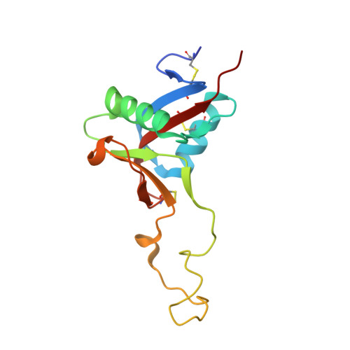
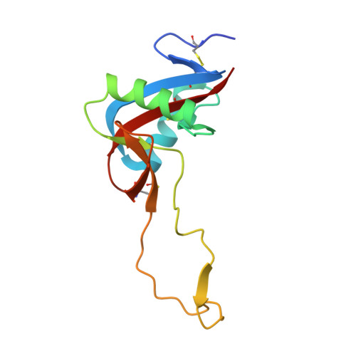
Structure of the Snake-Venom Toxin Convulxin

Batuwangala, T.Leduc, M.Gibbins, J.M.Bon, C.Jones, E.Y.

(2004) Acta Crystallogr D Biol Crystallogr 60: 46

  • DOI: https://doi.org/10.1107/s0907444903021620
  • Primary Citation of Related Structures:  
    1UOS

  • PubMed Abstract: 

    Snake venoms contain a number of proteins that interact with components of the haemostatic system that promote or inhibit events leading to blood-clot formation. The snake-venom protein convulxin (Cvx) binds glycoprotein (GP) VI, the platelet receptor for collagen, and triggers signal transduction. Here, the 2.7 A resolution crystal structure of Cvx is presented. In common with other members of this snake-venom protein family, Cvx is an alphabeta-heterodimer and conforms to the C-type lectin-fold topology. Comparison with other family members allows a set of Cvx residues that form a concave surface to be putatively implicated in GPVI binding. Unlike other family members, with the exception of flavocetin-A (FL-A), Cvx forms an (alphabeta)(4) tetramer. This oligomeric structure is consistent with Cvx clustering GPVI molecules on the surface of platelets and as a result promoting signal transduction activity. The Cvx structure and the location of the putative binding sites suggest a model for this multimeric signalling assembly.


  • Organizational Affiliation

    Cancer Research UK Receptor Structure Group, Division of Structural Biology, University of Oxford, The Henry Wellcome Building for Genomic Medicine, Roosevelt Drive, Headington, Oxford OX3 7BN, England.


Macromolecules
Find similar proteins by:  (by identity cutoff)  |  3D Structure
Entity ID: 1
MoleculeChains Sequence LengthOrganismDetailsImage
CONVULXIN ALPHA
A, C
135Crotalus durissus terrificusMutation(s): 0 
UniProt
Find proteins for O93426 (Crotalus durissus terrificus)
Explore O93426 
Go to UniProtKB:  O93426
Entity Groups  
Sequence Clusters30% Identity50% Identity70% Identity90% Identity95% Identity100% Identity
UniProt GroupO93426
Sequence Annotations
Expand
  • Reference Sequence
Find similar proteins by:  (by identity cutoff)  |  3D Structure
Entity ID: 2
MoleculeChains Sequence LengthOrganismDetailsImage
CONVULXIN BETA
B, D
126Crotalus durissus terrificusMutation(s): 0 
UniProt
Find proteins for O93427 (Crotalus durissus terrificus)
Explore O93427 
Go to UniProtKB:  O93427
Entity Groups  
Sequence Clusters30% Identity50% Identity70% Identity90% Identity95% Identity100% Identity
UniProt GroupO93427
Sequence Annotations
Expand
  • Reference Sequence
Experimental Data & Validation

Experimental Data

  • Method: X-RAY DIFFRACTION
  • Resolution: 2.70 Å
  • R-Value Free: 0.263 
  • R-Value Work: 0.235 
  • R-Value Observed: 0.235 
  • Space Group: I 4
Unit Cell:
Length ( Å )Angle ( ˚ )
a = 132.458α = 90
b = 132.458β = 90
c = 112.959γ = 90
Software Package:
Software NamePurpose
CNSrefinement
DENZOdata reduction
SCALEPACKdata scaling
CNSphasing

Structure Validation

View Full Validation Report


Entry History 

Deposition Data

Revision History  (Full details and data files)

  • Version 1.0: 2003-10-14
    Type: Initial release
  • Version 1.1: 2011-05-08
    Changes: Version format compliance
  • Version 1.2: 2011-07-13
    Changes: Version format compliance
  • Version 1.3: 2023-12-13
    Changes: Data collection, Database references, Other, Refinement description