A molecular mechanism for autoinhibition of the tandem SH3 domains of p47phox, the regulatory subunit of the phagocyte NADPH oxidase
Yuzawa, S., Suzuki, N.N., Fujioka, Y., Ogura, K., Sumimoto, H., Inagaki, F.(2004) Genes Cells 9: 443-456
- PubMed: 15147273
- DOI: https://doi.org/10.1111/j.1356-9597.2004.00733.x
- Primary Citation of Related Structures:
1UEC - PubMed Abstract:
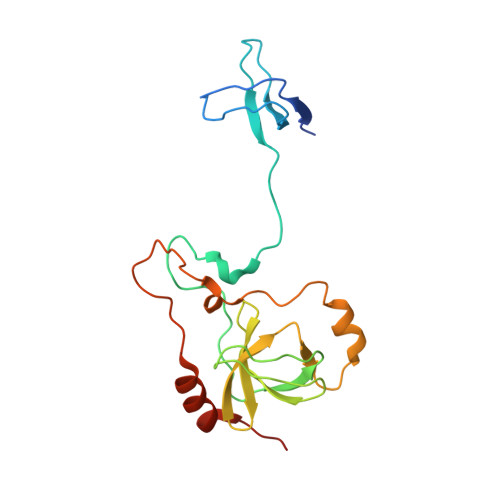
The phagocyte NADPH oxidase is a multisubunit enzyme responsible for the production of reactive oxygen species. p47(phox) is a cytosolic component of the NADPH oxidase and plays an important role in the assembly of the activated complex. The structural determination of the tandem SH3 domains of p47(phox) is crucial for elucidation of the molecular mechanism of the activation of p47(phox). We determined the X-ray crystal structure of the tandem SH3 domains with the polybasic/autoinhibitory region (PBR/AIR) of p47(phox). The GAPPR sequence involved in PBR/AIR forms a left-handed polyproline type-II helix (PPII) and interacts with the conserved SH3 binding surfaces of the SH3 domains simultaneously. These SH3 domains are related by a 2-fold pseudosymmetry axis at the centre of the binding groove and interact with the single PPII helix formed by the GAPPR sequence with opposite orientation. In addition, a number of intra-molecular interactions among the SH3 domains, PBR/AIR and the linker tightly hold the architecture of the tandem SH3 domains into the compact structure and stabilize the autoinhibited form synergistically. Phosphorylation of the serine residues in PBR/AIR could destabilize and successively release the intra-molecular interactions. Thus, the overall structure could be rearranged from the autoinhibitory conformation to the active conformation and the PPII ligand binding surfaces on the SH3 domains are now unmasked, which enables their interaction with the target sequence in p22(phox).
Organizational Affiliation:
Department of Structural Biology, Graduate School of Pharmaceutical Sciences, Hokkaido University, Sapporo 060-0812, Japan.