1U7T

Crystal Structure of ABAD/HSD10 with a Bound Inhibitor


Experimental Data Snapshot

  • Method: X-RAY DIFFRACTION
  • Resolution: 2.00 Å
  • R-Value Free: 0.263 
  • R-Value Work: 0.215 
  • R-Value Observed: 0.215 

wwPDB Validation   3D Report Full Report


This is version 1.6 of the entry. See complete history


Literature

Crystal structure of human ABAD/HSD10 with a bound inhibitor: implications for design of Alzheimer's disease therapeutics

Kissinger, C.R.Rejto, P.A.Pelletier, L.A.Thomson, J.A.Showalter, R.E.Abreo, M.A.Agree, C.S.Margosiak, S.Meng, J.J.Aust, R.M.Vanderpool, D.Li, B.Tempczyk-Russell, A.Villafranca, J.E.

(2004) J Mol Biol 342: 943-952

  • DOI: https://doi.org/10.1016/j.jmb.2004.07.071
  • Primary Citation of Related Structures:  
    1U7T

  • PubMed Abstract: 

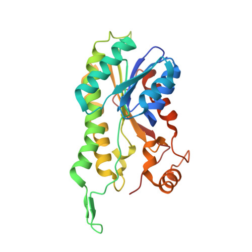
    The enzyme 17beta-hydroxysteroid dehydrogenase type 10 (HSD10), also known as amyloid beta-peptide-binding alcohol dehydrogenase (ABAD), has been implicated in the development of Alzheimer's disease. This protein, a member of the short-chain dehydrogenase/reductase family of enzymes, has been shown to bind beta-amyloid and to participate in beta-amyloid neurotoxicity. We have determined the crystal structure of human ABAD/HSD10 complexed with NAD(+) and an inhibitory small molecule. The inhibitor occupies the substrate-binding site and forms a covalent adduct with the NAD(+) cofactor. The crystal structure provides a basis for the design of potent, highly specific ABAD/HSD10 inhibitors with potential application in the treatment of Alzheimer's disease.


  • Organizational Affiliation

    Pfizer-La Jolla, 10777 Science Center Dr., San Diego, CA 92121, USA.


Macromolecules
Find similar proteins by:  (by identity cutoff)  |  3D Structure
Entity ID: 1
MoleculeChains Sequence LengthOrganismDetailsImage
3-hydroxyacyl-CoA dehydrogenase type II
A, B, C, D
261Homo sapiensMutation(s): 1 
Gene Names: HADH2ERABXH98G2SCHAD
EC: 1.1.1.35
UniProt & NIH Common Fund Data Resources
Find proteins for Q99714 (Homo sapiens)
Explore Q99714 
Go to UniProtKB:  Q99714
PHAROS:  Q99714
GTEx:  ENSG00000072506 
Entity Groups  
Sequence Clusters30% Identity50% Identity70% Identity90% Identity95% Identity100% Identity
UniProt GroupQ99714
Sequence Annotations
Expand
  • Reference Sequence
Small Molecules
Ligands 2 Unique
IDChains Name / Formula / InChI Key2D Diagram3D Interactions
TDT
Query on TDT

Download Ideal Coordinates CCD File 
E [auth A],
G [auth C],
H [auth D]
1-AZEPAN-1-YL-2-PHENYL-2-(4-THIOXO-1,4-DIHYDRO-PYRAZOLO[3,4-D]PYRIMIDIN-5-YL)ETHANONE ADDUCT
C40 H44 N12 O15 P2 S
XHBACRZQFHLVAV-LFFSAXSQSA-L
NAD
Query on NAD

Download Ideal Coordinates CCD File 
F [auth B]NICOTINAMIDE-ADENINE-DINUCLEOTIDE
C21 H27 N7 O14 P2
BAWFJGJZGIEFAR-NNYOXOHSSA-N
Binding Affinity Annotations 
IDSourceBinding Affinity
NAD Binding MOAD:  1U7T Ki: 4.25e+5 (nM) from 1 assay(s)
TDT Binding MOAD:  1U7T IC50: 92 (nM) from 1 assay(s)
Experimental Data & Validation

Experimental Data

  • Method: X-RAY DIFFRACTION
  • Resolution: 2.00 Å
  • R-Value Free: 0.263 
  • R-Value Work: 0.215 
  • R-Value Observed: 0.215 
  • Space Group: C 1 2 1
Unit Cell:
Length ( Å )Angle ( ˚ )
a = 122α = 90
b = 80.8β = 105.6
c = 110γ = 90
Software Package:
Software NamePurpose
DENZOdata reduction
SCALEPACKdata scaling
EPMRphasing
SHELXL-97refinement

Structure Validation

View Full Validation Report


Entry History 

Deposition Data

Revision History  (Full details and data files)

  • Version 1.0: 2004-10-05
    Type: Initial release
  • Version 1.1: 2008-04-30
    Changes: Version format compliance
  • Version 1.2: 2011-07-13
    Changes: Version format compliance
  • Version 1.3: 2018-04-04
    Changes: Data collection
  • Version 1.4: 2021-10-20
    Changes: Database references, Derived calculations
  • Version 1.5: 2024-02-14
    Changes: Data collection
  • Version 1.6: 2024-04-03
    Changes: Refinement description