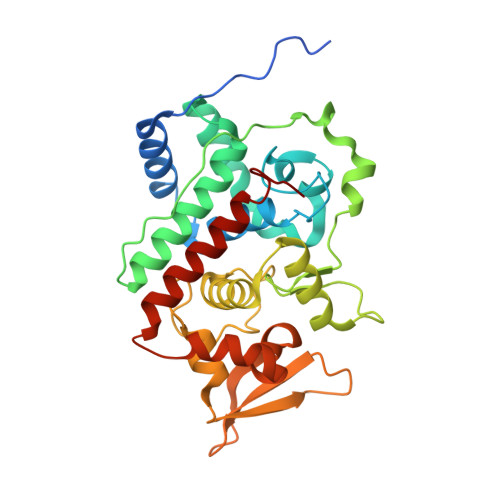
Structure of the C-terminal domain of the catalase-peroxidase KatG from Escherichia coli.
Carpena, X., Melik-Adamyan, W., Loewen, P.C., Fita, I.(2004) Acta Crystallogr D Biol Crystallogr 60: 1824-1832
- PubMed: 15388929
- DOI: https://doi.org/10.1107/S0907444904020621
- Primary Citation of Related Structures:
1U2J, 1U2K, 1U2L - PubMed Abstract:
Catalase-peroxidases or KatGs, the apparent in vivo activators of the anti-tubercular pro-drug isoniazid, are active as homodimers, each subunit having two distinct but sequence- and structure-related domains. The N-terminal domain contains the haem group and is catalytically active, while the C-terminal domain lacks the cofactor. The C-terminal domain of KatG from Escherichia coli is expressed as a soluble protein which has been crystallized in triclinic, orthorhombic and tetragonal crystal forms. Packing in the orthorhombic crystals, with eight molecules in the asymmetric unit, follows the pattern of commensurate modulated structures, which explains the diversity of pseudo-origin peaks observed in the native Patterson map. The different crystal forms arise from variations in the length and sequence of the N-terminal extensions in the different constructs. Despite the variability in the N-terminal region, the overall domain conformations beginning with Pro437 are very similar both to each other and to the C-terminal domains within the native structures of the KatGs from Haloarcula marismortui and Burkholderia pseudomallei. Some structural reorganization in the C-terminal domain relative to the N-terminal domain has evolved to compensate for the absence of the haem group. A high percentage of the residues in the C-terminal domains of KatG proteins from different sources are highly conserved and these residues are spread uniformly throughout the domain. The easily folded nature and retention of structure in the C-terminal domain suggests that it may serve as a platform for the folding of the N-terminal domain and for stabilization of the molecular dimer.
Organizational Affiliation:
Department of Microbiology, University of Manitoba, Winnipeg, MB, R3T 2N2, Canada.