1U06

crystal structure of chicken alpha-spectrin SH3 domain


Experimental Data Snapshot

  • Method: X-RAY DIFFRACTION
  • Resolution: 1.49 Å
  • R-Value Free: 0.189 
  • R-Value Work: 0.175 
  • R-Value Observed: 0.176 

wwPDB Validation   3D Report Full Report


This is version 1.4 of the entry. See complete history


Literature

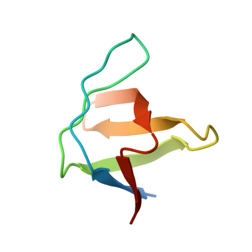
Detection of dynamic water molecules in a microcrystalline sample of the SH3 domain of alpha-spectrin by MAS solid-state NMR.

Chevelkov, V.Faelber, K.Diehl, A.Heinemann, U.Oschkinat, H.Reif, B.

(2005) J Biomol NMR 31: 295-310

  • DOI: https://doi.org/10.1007/s10858-005-1718-z
  • Primary Citation of Related Structures:  
    1U06

  • PubMed Abstract: 

    Water molecules are a major determinant of protein stability and are important for understanding protein-protein interactions. We present two experiments which allow to measure first the effective T(2) decay rate of individual amide proton, and second the magnetization build-up rates for a selective transfer from H(2)O to H(N) using spin diffusion as a mixing element. The experiments are demonstrated for a uniformly (2)H, (15)N labeled sample of a microcrystalline SH3 domain in which exchangeable deuterons were back-substituted with protons. In order to evaluate the NMR experimental data, as X-ray structure of the protein was determined using the same crystallization protocol as for the solid-state NMR sample. The NMR experimental data are correlated with the dipolar couplings calculated from H(2)O-H(N) distances which were extracted from the X-ray structure of the protein. We find that the H(N) T(2) decay rates and H(2)O-H(N) build-up rates are sensitive to distance and dynamics of the detected water molecules with respect to the protein. We show that qualitative information about localization and dynamics of internal water molecules can be obtained in the solid-state by interpretation of the spin dynamics of a reporter amide proton.


  • Organizational Affiliation

    Forschungsinstitut für Molekulare Pharmakologie (FMP), Berlin, Germany.


Macromolecules
Find similar proteins by:  (by identity cutoff)  |  3D Structure
Entity ID: 1
MoleculeChains Sequence LengthOrganismDetailsImage
Spectrin alpha chain, brain62Gallus gallusMutation(s): 0 
UniProt
Find proteins for P07751 (Gallus gallus)
Explore P07751 
Go to UniProtKB:  P07751
Entity Groups  
Sequence Clusters30% Identity50% Identity70% Identity90% Identity95% Identity100% Identity
UniProt GroupP07751
Sequence Annotations
Expand
  • Reference Sequence
Experimental Data & Validation

Experimental Data

  • Method: X-RAY DIFFRACTION
  • Resolution: 1.49 Å
  • R-Value Free: 0.189 
  • R-Value Work: 0.175 
  • R-Value Observed: 0.176 
  • Space Group: P 21 21 21
Unit Cell:
Length ( Å )Angle ( ˚ )
a = 33.6α = 90
b = 42.25β = 90
c = 49.61γ = 90
Software Package:
Software NamePurpose
MAR345data collection
XDSdata reduction
AMoREphasing
REFMACrefinement
XDSdata scaling

Structure Validation

View Full Validation Report


Entry History 

Deposition Data

Revision History  (Full details and data files)

  • Version 1.0: 2005-01-13
    Type: Initial release
  • Version 1.1: 2008-04-30
    Changes: Version format compliance
  • Version 1.2: 2011-07-13
    Changes: Version format compliance
  • Version 1.3: 2017-10-11
    Changes: Refinement description
  • Version 1.4: 2023-10-25
    Changes: Data collection, Database references, Derived calculations, Refinement description