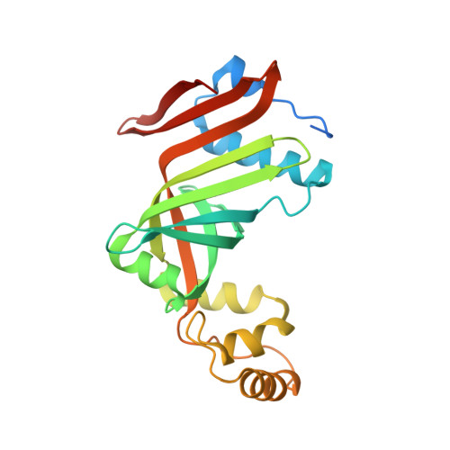
Structure of the phenazine biosynthesis enzyme PhzG.
Parsons, J.F., Calabrese, K., Eisenstein, E., Ladner, J.E.(2004) Acta Crystallogr D Biol Crystallogr 60: 2110-2113
- PubMed: 15502343
- DOI: https://doi.org/10.1107/S0907444904022474
- Primary Citation of Related Structures:
1T9M, 1TY9 - PubMed Abstract:
PhzG is a flavin-dependent oxidase that is believed to play a role in phenazine antibiotic synthesis in various bacteria, including Pseudomonas. Phenazines are chorismic acid derivatives that provide the producing organisms, including the opportunistic pathogen P. aeruginosa, with a competitive growth advantage. Here, the crystal structures of PhzG from both P. aeruginosa and P. fluorescens solved in an unliganded state at 1.9 and 1.8 A resolution, respectively, are described. Although the specific reaction in phenazine biosynthesis catalyzed by PhzG is unknown, the structural data indicates that PhzG is closely related to pyridoxine-5'-phosphate oxidase, the Escherichia coli pdxH gene product, which catalyzes the final step in pyridoxal-5'-phosphate (PLP) biosynthesis. A previous proposal suggested that the physiological substrate of PhzG to be 2,3-dihydro-3-hydroxyanthranilic acid (DHHA), a phenazine precursor produced by the sequential actions of the PhzE and PhzD enzymes on chorismate, and that two DHHA molecules dimerized in another enzyme-catalyzed reaction to yield phenazine-1-carboxylate. However, it was not possible to demonstrate any in vitro activity upon incubation of PhzG and DHHA. Interestingly, analysis of the in vitro activities of PhzG in combination with PhzF suggests that PhzF acts on DHHA and that PhzG then reacts with a non-aromatic tricyclic phenazine precusor to catalyze an oxidation/aromatization reaction that yields phenazine-1-carboxylate. It is proposed that phzG arose by duplication of pdxH and that the subtle differences seen between the structures of PhzG and PdxH correlate with the loss of the ability of PhzG to catalyze PLP formation. Sequence alignments and superimpositions of the active sites of PhzG and PdxH reveal that the residues that form a positively charged pocket around the phosphate of PLP in the PdxH-PLP complex are not conserved in PhzG, consistent with the inability of phosphorylated compounds to serve as substrates for PhzG.
Organizational Affiliation:
Center for Advanced Research in Biotechnology, University of Maryland Biotechnology Institute, National Institute of Standards and Technology, 9600 Gudelsky Drive, Rockville, MD 20850, USA.