1TMX

Crystal structure of hydroxyquinol 1,2-dioxygenase from Nocardioides Simplex 3E


Experimental Data Snapshot

  • Method: X-RAY DIFFRACTION
  • Resolution: 1.75 Å
  • R-Value Free: 0.247 
  • R-Value Work: 0.193 
  • R-Value Observed: 0.195 

wwPDB Validation   3D Report Full Report


Ligand Structure Quality Assessment 


This is version 1.4 of the entry. See complete history


Literature

Crystal structure of the hydroxyquinol 1,2-dioxygenase from Nocardioides simplex 3E, a key enzyme involved in polychlorinated aromatics biodegradation.

Ferraroni, M.Seifert, J.Travkin, V.M.Thiel, M.Kaschabek, S.Scozzafava, A.Golovleva, L.Schlomann, M.Briganti, F.

(2005) J Biol Chem 280: 21144-21154

  • DOI: https://doi.org/10.1074/jbc.M500666200
  • Primary Citation of Related Structures:  
    1TMX

  • PubMed Abstract: 

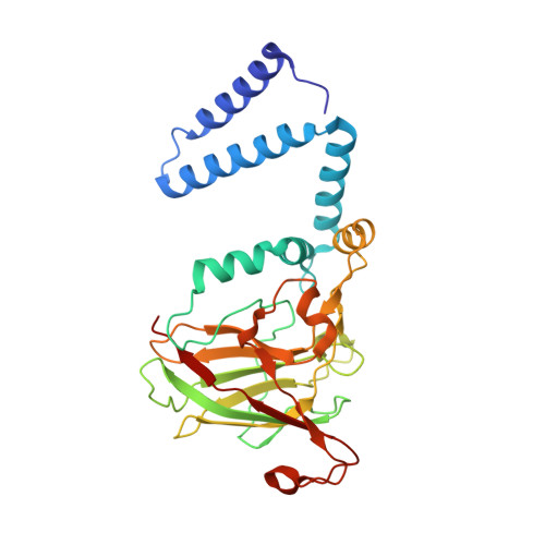
    Hydroxyquinol 1,2-dioxygenase (1,2-HQD) catalyzes the ring cleavage of hydroxyquinol (1,2,4-trihydroxybenzene), a central intermediate in the degradation of aromatic compounds including a variety of particularly recalcitrant polychloro- and nitroaromatic pollutants. We report here the primary sequence determination and the analysis of the crystal structure of the 1,2-HQD from Nocardioides simplex 3E solved at 1.75 A resolution using the multiple wavelength anomalous dispersion of the two catalytic irons (1 Fe/293 amino acids). The catalytic Fe(III) coordination polyhedron composed by the side chains of Tyr164, Tyr197, His221, and His223 resembles that of the other known intradiol-cleaving dioxygenases, but several of the tertiary structure features are notably different. One of the most distinctive characteristics of the present structure is the extensive openings and consequent exposure to solvent of the upper part of the catalytic cavity arranged to favor the binding of hydroxyquinols but not catechols. A co-crystallized benzoate-like molecule is also found bound to the metal center forming a distinctive hydrogen bond network as observed previously also in 4-chlorocatechol 1,2-dioxygenase from Rhodococcus opacus 1CP. This is the first structure of an intradiol dioxygenase specialized in hydroxyquinol ring cleavage to be investigated in detail.


  • Organizational Affiliation

    Dipartimento di Chimica, Università di Firenze, Via della Lastruccia 3, Sesto Fiorentino I-50019, Italy.


Macromolecules
Find similar proteins by:  (by identity cutoff)  |  3D Structure
Entity ID: 1
MoleculeChains Sequence LengthOrganismDetailsImage
hydroxyquinol 1,2-dioxygenase
A, B
293Pimelobacter simplexMutation(s): 0 
EC: 1.13.11.37
UniProt
Find proteins for Q5PXQ6 (Nocardioides simplex)
Explore Q5PXQ6 
Go to UniProtKB:  Q5PXQ6
Entity Groups  
Sequence Clusters30% Identity50% Identity70% Identity90% Identity95% Identity100% Identity
UniProt GroupQ5PXQ6
Sequence Annotations
Expand
  • Reference Sequence
Small Molecules
Ligands 6 Unique
IDChains Name / Formula / InChI Key2D Diagram3D Interactions
HGX
Query on HGX

Download Ideal Coordinates CCD File 
G [auth A],
K [auth B]
1-HEPTADECANOYL-2-TRIDECANOYL-3-GLYCEROL-PHOSPHONYL CHOLINE
C38 H77 N O8 P
AZEAMUNCULVBPK-PSXMRANNSA-O
BEZ
Query on BEZ

Download Ideal Coordinates CCD File 
H [auth A],
L [auth B]
BENZOIC ACID
C7 H6 O2
WPYMKLBDIGXBTP-UHFFFAOYSA-N
SO4
Query on SO4

Download Ideal Coordinates CCD File 
F [auth A],
J [auth B]
SULFATE ION
O4 S
QAOWNCQODCNURD-UHFFFAOYSA-L
CU
Query on CU

Download Ideal Coordinates CCD File 
D [auth A]COPPER (II) ION
Cu
JPVYNHNXODAKFH-UHFFFAOYSA-N
FE
Query on FE

Download Ideal Coordinates CCD File 
C [auth A],
I [auth B]
FE (III) ION
Fe
VTLYFUHAOXGGBS-UHFFFAOYSA-N
CL
Query on CL

Download Ideal Coordinates CCD File 
E [auth A]CHLORIDE ION
Cl
VEXZGXHMUGYJMC-UHFFFAOYSA-M
Experimental Data & Validation

Experimental Data

  • Method: X-RAY DIFFRACTION
  • Resolution: 1.75 Å
  • R-Value Free: 0.247 
  • R-Value Work: 0.193 
  • R-Value Observed: 0.195 
  • Space Group: P 1 21 1
Unit Cell:
Length ( Å )Angle ( ˚ )
a = 46.283α = 90
b = 84.978β = 92.84
c = 83.923γ = 90
Software Package:
Software NamePurpose
REFMACrefinement
MAR345data collection
SCALEPACKdata scaling
SOLVEphasing

Structure Validation

View Full Validation Report


Ligand Structure Quality Assessment 


Entry History 

Deposition Data

Revision History  (Full details and data files)

  • Version 1.0: 2005-03-22
    Type: Initial release
  • Version 1.1: 2008-04-30
    Changes: Version format compliance
  • Version 1.2: 2011-07-13
    Changes: Version format compliance
  • Version 1.3: 2017-10-11
    Changes: Refinement description
  • Version 1.4: 2024-02-14
    Changes: Data collection, Database references, Derived calculations