1T2L

Three Crystal Structures of Human Coactosin-like Protein


Experimental Data Snapshot

  • Method: X-RAY DIFFRACTION
  • Resolution: 2.80 Å
  • R-Value Free: 0.269 
  • R-Value Work: 0.197 
  • R-Value Observed: 0.205 

wwPDB Validation   3D Report Full Report


This is version 1.2 of the entry. See complete history


Literature

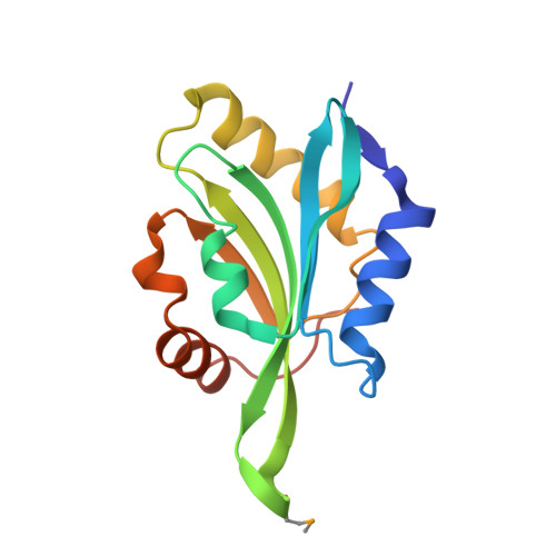
Crystal Structure of Human Coactosin-like Protein

Liu, L.Wei, Z.Wang, Y.Wan, M.Cheng, Z.Gong, W.

(2004) J Mol Biol 344: 317-323

  • DOI: https://doi.org/10.1016/j.jmb.2004.09.036
  • Primary Citation of Related Structures:  
    1T2L

  • PubMed Abstract: 

    Human coactosin-like protein is an actin filament binding protein but does not bind to globular actin. It associates with 5-Lipoxygenase both in vivo and in vitro, playing important roles in modulating the activities of actin and 5-Lipoxygenase. Coactosin counteracts the capping activity of capping protein which inhibits the actin polymerization. We determined the crystal structures of human coactosin-like protein by multi-wavelength anomalous dispersion method. The structure showed a high level of similarity to ADF-H domain, although their amino acid sequences share low degree of homology. A few conserved hydrophobic residues that may contribute to the folding were identified. This structure suggests coactosin-like protein bind to F-actin in a different way from ADF/Cofilin family. Combined with the information from previous mutagenesis studies, the binding sites for F-actin and 5-Lipoxygenase were analyzed, respectively. These two sites are quite close, which might prevent F-actin and 5-Lipoxygenase from binding to coactosin simultaneously.


  • Organizational Affiliation

    National Laboratory of Biomacromolecules, Institute of Biophysics, Chinese Academy of Sciences, Beijing 100101, People's Republic of China.


Macromolecules
Find similar proteins by:  (by identity cutoff)  |  3D Structure
Entity ID: 1
MoleculeChains Sequence LengthOrganismDetailsImage
Coactosin-like protein
A, B
141Homo sapiensMutation(s): 1 
UniProt & NIH Common Fund Data Resources
Find proteins for Q14019 (Homo sapiens)
Explore Q14019 
Go to UniProtKB:  Q14019
PHAROS:  Q14019
GTEx:  ENSG00000103187 
Entity Groups  
Sequence Clusters30% Identity50% Identity70% Identity90% Identity95% Identity100% Identity
UniProt GroupQ14019
Sequence Annotations
Expand
  • Reference Sequence
Small Molecules
Modified Residues  1 Unique
IDChains TypeFormula2D DiagramParent
MSE
Query on MSE
A, B
L-PEPTIDE LINKINGC5 H11 N O2 SeMET
Experimental Data & Validation

Experimental Data

  • Method: X-RAY DIFFRACTION
  • Resolution: 2.80 Å
  • R-Value Free: 0.269 
  • R-Value Work: 0.197 
  • R-Value Observed: 0.205 
  • Space Group: C 1 2 1
Unit Cell:
Length ( Å )Angle ( ˚ )
a = 124.018α = 90
b = 37.033β = 106.83
c = 60.322γ = 90
Software Package:
Software NamePurpose
DENZOdata reduction
SCALEPACKdata scaling
SOLVEphasing
CNSrefinement

Structure Validation

View Full Validation Report


Entry History 

Deposition Data

Revision History  (Full details and data files)

  • Version 1.0: 2004-11-23
    Type: Initial release
  • Version 1.1: 2008-04-30
    Changes: Version format compliance
  • Version 1.2: 2011-07-13
    Changes: Version format compliance