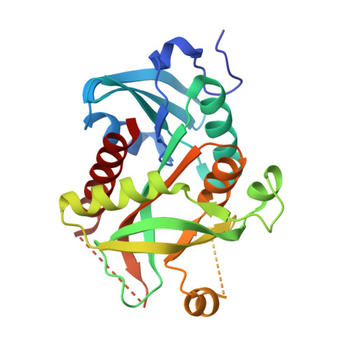
Crystal structures of escherichia coli uridine phosphorylase in two native and three complexed forms reveal basis of substrate specificity, induced conformational changes and influence of potassium
Caradoc-Davies, T.T., Cutfield, S.M., Lamont, I.L., Cutfield, J.F.(2004) J Mol Biol 337: 337-354
- PubMed: 15003451
- DOI: https://doi.org/10.1016/j.jmb.2004.01.039
- Primary Citation of Related Structures:
1RXC, 1RXS, 1RXU, 1RXY, 1T0U - PubMed Abstract:
Uridine phosphorylase (UP) is a key enzyme in the pyrimidine salvage pathway that catalyses the reversible phosphorolysis of uridine to uracil and ribose 1-phosphate. Inhibiting liver UP in humans raises blood uridine levels and produces a protective effect ("uridine rescue") against the toxicity of the chemotherapeutic agent 5-fluorouracil without reducing its antitumour activity. We have investigated UP-substrate interactions by determining the crystal structures of native Escherichia coli UP (two forms), and complexes with 5-fluorouracil/ribose 1-phosphate, 2-deoxyuridine/phosphate and thymidine/phosphate. These hexameric structures confirm the overall structural similarity of UP to E.coli purine nucleoside phosphorylase (PNP) whereby, in the presence of substrate, each displays a closed conformation resulting from a concerted movement that closes the active site cleft. However, in contrast to PNP where helix segmentation is the major conformational change between the open and closed forms, in UP more extensive changes are observed. In particular a swinging movement of a flap region consisting of residues 224-234 seals the active site. This overall change in conformation results in compression of the active site cleft. Gln166 and Arg168, part of an inserted segment not seen in PNP, are key residues in the uracil binding pocket and together with a tightly bound water molecule are seen to be involved in the substrate specificity of UP. Enzyme activity shows a twofold dependence on potassium ion concentration. The presence of a potassium ion at the monomer/monomer interface induces some local rearrangement, which results in dimer stabilisation. The conservation of key residues and interactions with substrate in the phosphate and ribose binding pockets suggest that ribooxocarbenium ion formation during catalysis of UP may be similar to that proposed for E.coli PNP.
Organizational Affiliation:
Department of Biochemistry, Otago School of Medical Sciences, University of Otago, P.O. Box 56, Dunedin 9001, New Zealand. tom.caradoc-davies@stonebow.otago.ac.nz