1SVY

SEVERIN DOMAIN 2, 1.75 ANGSTROM CRYSTAL STRUCTURE


Experimental Data Snapshot

  • Method: X-RAY DIFFRACTION
  • Resolution: 1.75 Å
  • R-Value Free: 0.246 
  • R-Value Work: 0.184 
  • R-Value Observed: 0.184 

wwPDB Validation   3D Report Full Report


This is version 1.2 of the entry. See complete history


Literature

Mapping the functional surface of domain 2 in the gelsolin superfamily.

Puius, Y.A.Fedorov, E.V.Eichinger, L.Schleicher, M.Almo, S.C.

(2000) Biochemistry 39: 5322-5331

  • DOI: https://doi.org/10.1021/bi992364d
  • Primary Citation of Related Structures:  
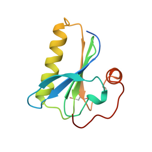
    1SVY

  • PubMed Abstract: 

    The crystal structure of the F-actin binding domain 2 of severin, the gelsolin homologue from Dictyostelium discoideum, has been determined by multiple isomorphous replacement and refined to 1.75 A resolution. The structure reveals an alpha-helix-beta-sheet sandwich similar to the domains of gelsolin and villin, and contains two cation-binding sites, as observed in other domain 1 and domain 2 homologues. Comparison of the structures of several gelsolin family domains has identified residues that may mediate F-actin binding in gelsolin domain 2 homologues. To assess the involvement of these residues in F-actin binding, three mutants of human gelsolin domain 2 were assayed for F-actin binding activity and thermodynamic stability. Two of the mutants, RRV168AAA and RLK210AAA, demonstrated a lowered affinity for F-actin, indicating a role for those residues in filament binding. Using both structural and biochemical data, we have constructed a model of the gelsolin domain 1-domain 2-F-actin complex. This model highlights a number of interactions that may serve as positive and negative determinants of filament end- and side-binding.


  • Organizational Affiliation

    Department of Biochemistry, Albert Einstein College of Medicine, 1300 Morris Park Avenue, Bronx, New York 10461, USA.


Macromolecules
Find similar proteins by:  (by identity cutoff)  |  3D Structure
Entity ID: 1
MoleculeChains Sequence LengthOrganismDetailsImage
SEVERIN114Dictyostelium discoideumMutation(s): 0 
UniProt
Find proteins for P10733 (Dictyostelium discoideum)
Explore P10733 
Go to UniProtKB:  P10733
Entity Groups  
Sequence Clusters30% Identity50% Identity70% Identity90% Identity95% Identity100% Identity
UniProt GroupP10733
Sequence Annotations
Expand
  • Reference Sequence
Small Molecules
Ligands 2 Unique
IDChains Name / Formula / InChI Key2D Diagram3D Interactions
CA
Query on CA

Download Ideal Coordinates CCD File 
B [auth A]CALCIUM ION
Ca
BHPQYMZQTOCNFJ-UHFFFAOYSA-N
NA
Query on NA

Download Ideal Coordinates CCD File 
C [auth A]SODIUM ION
Na
FKNQFGJONOIPTF-UHFFFAOYSA-N
Modified Residues  1 Unique
IDChains TypeFormula2D DiagramParent
CSO
Query on CSO
A
L-PEPTIDE LINKINGC3 H7 N O3 SCYS
Experimental Data & Validation

Experimental Data

  • Method: X-RAY DIFFRACTION
  • Resolution: 1.75 Å
  • R-Value Free: 0.246 
  • R-Value Work: 0.184 
  • R-Value Observed: 0.184 
  • Space Group: P 21 21 21
Unit Cell:
Length ( Å )Angle ( ˚ )
a = 33.352α = 90
b = 49.218β = 90
c = 62.79γ = 90
Software Package:
Software NamePurpose
PHASES-95model building
X-PLORmodel building
X-PLORrefinement
DENZOdata reduction
SCALEPACKdata scaling
PHASESphasing
X-PLORphasing

Structure Validation

View Full Validation Report


Entry History 

Deposition Data

Revision History  (Full details and data files)

  • Version 1.0: 1999-08-10
    Type: Initial release
  • Version 1.1: 2008-03-24
    Changes: Version format compliance
  • Version 1.2: 2011-07-13
    Changes: Version format compliance