1S6B

X-ray Crystal Structure of a Complex Formed Between Two Homologous Isoforms of Phospholipase A2 from Naja naja sagittifera: Principle of Molecular Association and Inactivation


Experimental Data Snapshot

  • Method: X-RAY DIFFRACTION
  • Resolution: 1.60 Å
  • R-Value Free: 0.236 
  • R-Value Work: 0.206 
  • R-Value Observed: 0.212 

wwPDB Validation   3D Report Full Report


This is version 1.4 of the entry. See complete history


Literature

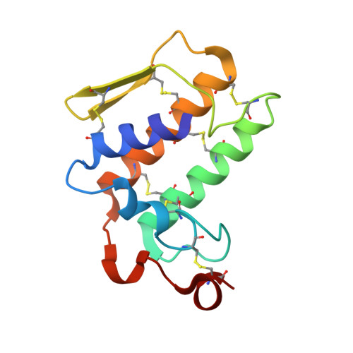
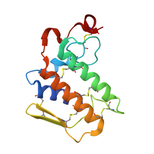
Crystal structure of a calcium-induced dimer of two isoforms of cobra phospholipase A2 at 1.6 A resolution.

Jabeen, T.Sharma, S.Singh, N.Singh, R.K.Kaur, P.Perbandt, M.Betzel, C.h.Srinivasan, A.Singh, T.P.

(2005) Proteins 59: 856-863

  • DOI: https://doi.org/10.1002/prot.20464
  • Primary Citation of Related Structures:  
    1S6B

  • PubMed Abstract: 

    The calcium-induced formation of a complex between two isoforms of cobra venom phospholipase A2 reveals a novel interplay between the monomer-dimer and activity-inactivity transitions. The monodispersed isoforms lack activity in the absence of calcium ions while both molecules gain activity in the presence of calcium ions. At concentrations higher than 10 mg/ml, in the presence of calcium ions, they dimerize and lose activity again. The present study reports the crystal structure of a calcium-induced dimer between two isoforms of cobra phospholipase A2. In the complex, one molecule contains a calcium ion in the calcium binding loop while the second molecule does not possess an intramolecular calcium ion. However, there are two calcium ions per dimer in the structure. The second calcium ion is present at an intermolecular site and that is presumably responsible for the dimerization. The calcium binding loops of the two molecules adopt strikingly different conformations. The so-called calcium binding loop in the calcium-containing molecule adopts a normal conformation as generally observed in other calcium containing phospholipase A(2) enzymes while the conformation of the corresponding loop in the calcium free monomer deviates considerably with the formation of a unique intraloop Gly33 (N)-Cys27 (O) = 2.74 A backbone hydrogen bond. The interactions of Arg31 (B) with Asp49 (A) and absence of calcium ion are responsible for the loss of catalytic activity in molecule A while interactions of Arg2 (B) with Tyr52 (B) inactivate molecule B.


  • Organizational Affiliation

    Department of Biophysics, All India Institute of Medical Sciences, New Delhi, India.


Macromolecules
Find similar proteins by:  (by identity cutoff)  |  3D Structure
Entity ID: 1
MoleculeChains Sequence LengthOrganismDetailsImage
Phospholipase A2 isoform 1119Naja sagittiferaMutation(s): 0 
EC: 3.1.1.4
UniProt
Find proteins for P60043 (Naja sagittifera)
Explore P60043 
Go to UniProtKB:  P60043
Entity Groups  
Sequence Clusters30% Identity50% Identity70% Identity90% Identity95% Identity100% Identity
UniProt GroupP60043
Sequence Annotations
Expand
  • Reference Sequence
Find similar proteins by:  (by identity cutoff)  |  3D Structure
Entity ID: 2
MoleculeChains Sequence LengthOrganismDetailsImage
Phospholipase A2 isoform 2119Naja sagittiferaMutation(s): 0 
EC: 3.1.1.4
UniProt
Find proteins for P60044 (Naja sagittifera)
Explore P60044 
Go to UniProtKB:  P60044
Entity Groups  
Sequence Clusters30% Identity50% Identity70% Identity90% Identity95% Identity100% Identity
UniProt GroupP60044
Sequence Annotations
Expand
  • Reference Sequence
Experimental Data & Validation

Experimental Data

  • Method: X-RAY DIFFRACTION
  • Resolution: 1.60 Å
  • R-Value Free: 0.236 
  • R-Value Work: 0.206 
  • R-Value Observed: 0.212 
  • Space Group: P 41
Unit Cell:
Length ( Å )Angle ( ˚ )
a = 64.537α = 90
b = 64.537β = 90
c = 57.117γ = 90
Software Package:
Software NamePurpose
DENZOdata reduction
SCALEPACKdata scaling
AMoREphasing
REFMACrefinement

Structure Validation

View Full Validation Report


Entry History 

Deposition Data

Revision History  (Full details and data files)

  • Version 1.0: 2004-02-10
    Type: Initial release
  • Version 1.1: 2008-04-29
    Changes: Version format compliance
  • Version 1.2: 2011-07-13
    Changes: Version format compliance
  • Version 1.3: 2018-02-07
    Changes: Experimental preparation
  • Version 1.4: 2023-10-25
    Changes: Data collection, Database references, Derived calculations, Refinement description