1RM0

Crystal Structure of Myo-Inositol 1-Phosphate Synthase From Saccharomyces cerevisiae In Complex With NAD+ and 2-deoxy-D-glucitol 6-(E)-vinylhomophosphonate


Experimental Data Snapshot

  • Method: X-RAY DIFFRACTION
  • Resolution: 2.05 Å
  • R-Value Free: 0.244 
  • R-Value Work: 0.189 
  • R-Value Observed: 0.189 

wwPDB Validation   3D Report Full Report


Ligand Structure Quality Assessment 


This is version 1.3 of the entry. See complete history


Literature

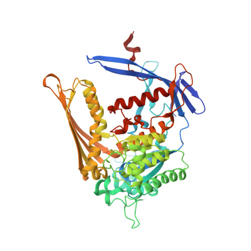
The structure of the 1L-myo-inositol-1-phosphate synthase-NAD+-2-deoxy-D-glucitol 6-(E)-vinylhomophosphonate complex demands a revision of the enzyme mechanism.

Jin, X.Foley, K.M.Geiger, J.H.

(2004) J Biol Chem 279: 13889-13895

  • DOI: https://doi.org/10.1074/jbc.M308986200
  • Primary Citation of Related Structures:  
    1RM0

  • PubMed Abstract: 

    1l-myo-inositol 1-phosphate (MIP) synthase catalyzes the conversion of d-glucose 6-phosphate to 1l-myo-inositol 1-phosphate, the first and rate-limiting step in the biosynthesis of all inositol-containing compounds. It involves an oxidation, enolization, intramolecular aldol cyclization, and reduction. Here we present the structure of MIP synthase in complex with NAD(+) and a high-affinity inhibitor, 2-deoxy-d-glucitol 6-(E)-vinylhomophosphonate. This structure reveals interactions between the enzyme active site residues and the inhibitor that are significantly different from that proposed for 2-deoxy-d-glucitol 6-phosphate in the previously published structure of MIP synthase-NAD(+)-2-deoxy-d-glucitol 6-phosphate. There are several other conformational changes in NAD(+) and the enzyme active site as well. Based on the new structural data, we propose a new and completely different mechanism for MIP synthase.


  • Organizational Affiliation

    Michigan State University, Chemistry Department, East Lansing, Michigan 48824, USA.


Macromolecules
Find similar proteins by:  (by identity cutoff)  |  3D Structure
Entity ID: 1
MoleculeChains Sequence LengthOrganismDetailsImage
myo-inositol-phosphate synthase
A, B
533Saccharomyces cerevisiaeMutation(s): 0 
Gene Names: INO1
EC: 5.5.1.4
UniProt
Find proteins for P11986 (Saccharomyces cerevisiae (strain ATCC 204508 / S288c))
Explore P11986 
Go to UniProtKB:  P11986
Entity Groups  
Sequence Clusters30% Identity50% Identity70% Identity90% Identity95% Identity100% Identity
UniProt GroupP11986
Sequence Annotations
Expand
  • Reference Sequence
Small Molecules
Binding Affinity Annotations 
IDSourceBinding Affinity
D6P Binding MOAD:  1RM0 Ki: 670 (nM) from 1 assay(s)
Experimental Data & Validation

Experimental Data

  • Method: X-RAY DIFFRACTION
  • Resolution: 2.05 Å
  • R-Value Free: 0.244 
  • R-Value Work: 0.189 
  • R-Value Observed: 0.189 
  • Space Group: C 1 2 1
Unit Cell:
Length ( Å )Angle ( ˚ )
a = 151.81α = 90
b = 97.78β = 126.3
c = 122.29γ = 90
Software Package:
Software NamePurpose
DENZOdata reduction
SCALEPACKdata scaling
CNSrefinement
CNSphasing

Structure Validation

View Full Validation Report


Ligand Structure Quality Assessment 


Entry History 

Deposition Data

Revision History  (Full details and data files)

  • Version 1.0: 2004-05-25
    Type: Initial release
  • Version 1.1: 2008-04-29
    Changes: Version format compliance
  • Version 1.2: 2011-07-13
    Changes: Derived calculations, Version format compliance
  • Version 1.3: 2024-02-14
    Changes: Data collection, Database references, Derived calculations