1R4F

Inosine-Adenosine-Guanosine Preferring Nucleoside Hydrolase From Trypanosoma vivax: Trp260Ala Mutant In Complex With 3-Deaza-Adenosine


Experimental Data Snapshot

  • Method: X-RAY DIFFRACTION
  • Resolution: 2.30 Å
  • R-Value Free: 0.257 
  • R-Value Work: 0.205 
  • R-Value Observed: 0.208 

wwPDB Validation   3D Report Full Report


Ligand Structure Quality Assessment 


This is version 1.4 of the entry. See complete history


Literature

Leaving group activation by aromatic stacking: an alternative to general Acid catalysis.

Versees, W.Loverix, S.Vandemeulebroucke, A.Geerlings, P.Steyaert, J.

(2004) J Mol Biol 338: 1-6

  • DOI: https://doi.org/10.1016/j.jmb.2004.02.049
  • Primary Citation of Related Structures:  
    1R4F

  • PubMed Abstract: 

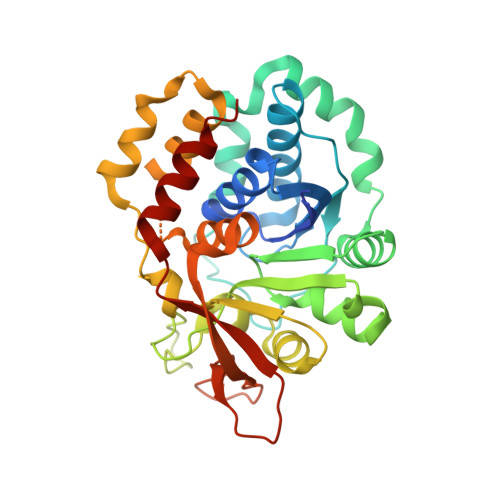
    General acid catalysis is a powerful and widely used strategy in enzymatic nucleophilic displacement reactions. For example, hydrolysis/phosphorolysis of the N-glycosidic bond in nucleosides and nucleotides commonly involves the protonation of the leaving nucleobase concomitant with nucleophilic attack. However, in the nucleoside hydrolase of the parasite Trypanosoma vivax, crystallographic and mutagenesis studies failed to identify a general acid. This enzyme binds the purine base of the substrate between the aromatic side-chains of Trp83 and Trp260. Here, we show via quantum chemical calculations that face-to-face stacking can raise the pKa of a heterocyclic aromatic compound by several units. Site-directed mutagenesis combined with substrate engineering demonstrates that Trp260 catalyzes the cleavage of the glycosidic bond by promoting the protonation of the purine base at N-7, hence functioning as an alternative to general acid catalysis.


  • Organizational Affiliation

    Laboratorium voor Ultrastructuur, Instituut voor Moleculaire Biologie, Vrije Universiteit Brussel and Vlaams Interuniversitair instituut voor Biotechnologie, Pleinlaan 2, 1050 Brussels, Belgium. wversees@vub.ac.be


Macromolecules
Find similar proteins by:  (by identity cutoff)  |  3D Structure
Entity ID: 1
MoleculeChains Sequence LengthOrganismDetailsImage
IAG-nucleoside hydrolase
A, B
339Trypanosoma vivaxMutation(s): 1 
EC: 3.2.2.1
UniProt
Find proteins for Q9GPQ4 (Trypanosoma vivax)
Explore Q9GPQ4 
Go to UniProtKB:  Q9GPQ4
Entity Groups  
Sequence Clusters30% Identity50% Identity70% Identity90% Identity95% Identity100% Identity
UniProt GroupQ9GPQ4
Sequence Annotations
Expand
  • Reference Sequence
Experimental Data & Validation

Experimental Data

  • Method: X-RAY DIFFRACTION
  • Resolution: 2.30 Å
  • R-Value Free: 0.257 
  • R-Value Work: 0.205 
  • R-Value Observed: 0.208 
  • Space Group: P 1 21 1
Unit Cell:
Length ( Å )Angle ( ˚ )
a = 51.996α = 90
b = 74.865β = 102.291
c = 81.369γ = 90
Software Package:
Software NamePurpose
DENZOdata reduction
SCALEPACKdata scaling
CNSrefinement
CNSphasing

Structure Validation

View Full Validation Report


Ligand Structure Quality Assessment 


Entry History 

Deposition Data

Revision History  (Full details and data files)

  • Version 1.0: 2004-04-13
    Type: Initial release
  • Version 1.1: 2008-04-29
    Changes: Version format compliance
  • Version 1.2: 2011-07-13
    Changes: Version format compliance
  • Version 1.3: 2021-10-27
    Changes: Database references, Derived calculations
  • Version 1.4: 2023-08-23
    Changes: Data collection, Refinement description