Crystal structure of the purine nucleoside phosphorylase (PNP) from Cellulomonas sp. and its implication for the mechanism of trimeric PNPs.
Tebbe, J., Bzowska, A., Wielgus-Kutrowska, B., Schroder, W., Kazimierczuk, Z., Shugar, D., Saenger, W., Koellner, G.(1999) J Mol Biol 294: 1239-1255
- PubMed: 10600382
- DOI: https://doi.org/10.1006/jmbi.1999.3327
- Primary Citation of Related Structures:
1C3X, 1QE5 - PubMed Abstract:
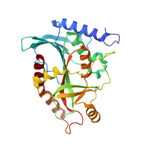
The three-dimensional structure of the trimeric purine nucleoside phosphorylase (PNP) from Cellulomonas sp. has been determined by X-ray crystallography. The binary complex of the enzyme with orthophosphate was crystallized in the orthorhombic space group P212121 with unit cell dimensions a=64.1 A, b=108.9 A, c=119.3 A and an enzymatically active trimer in the asymmetric unit. X-ray data were collected at 4 degrees C using synchrotron radiation (EMBL/DESY, Hamburg). The structure was solved by molecular replacement, with the calf spleen PNP structure as a model, and refined at 2.2 A resolution. The ternary "dead-end" complex of the enzyme with orthophosphate and 8-iodoguanine was obtained by soaking crystals of the binary orthophosphate complex with the very weak substrate 8-iodoguanosine. Data were collected at 100 K with CuKalpha radiation, and the three-dimensional structure refined at 2.4 A resolution. Although the sequence of the Cellulomonas PNP shares only 33 % identity with the calf spleen enzyme, and almost no identity with the hexameric Escherichia coli PNP, all three enzymes have many common structural features, viz. the nine-stranded central beta-sheet, the positions of the active centres, and the geometrical arrangement of the ligands in the active centres. Some similarities of the surrounding helices also prevail. In Cellulomonas PNP, each of the three active centres per trimer is occupied by orthophosphate, and by orthophosphate and base, respectively, and small structural differences between monomers A, B and C are observed. This supports cooperativity between subunits (non-identity of binding sites) rather than existence of more than one binding site per monomer, as previously suggested for binding of phosphate by mammalian PNPs. The phosphate binding site is located between two conserved beta- and gamma-turns and consists of Ser46, Arg103, His105, Gly135 and Ser223, and one or two water molecules. The guanine base is recognized by a zig-zag pattern of possible hydrogen bonds, as follows: guanine N-1...Glu204 O(epsilon1)...guanine NH2...Glu204 O(epsilon2). The exocyclic O6 of the base is bridged via a water molecule to Asn246 N(delta), which accounts for the inhibitory, but lack of substrate, activity of adenosine. An alternative molecular mechanism for catalysis by trimeric PNPs is proposed, in which the key catalytic role is played by Glu204 (Glu201 in the calf and human enzymes), while Asn246 (Asn243 in the mammalian enzymes) supports binding of 6-oxopurines rather than catalysis. This mechanism, in contrast to that previously suggested, is consistent with the excellent substrate properties of N-7 substituted nucleosides, the specificity of trimeric PNPs versus 6-oxopurine nucleosides and the reported kinetic properties of Glu201/Ala and Asn243/Ala point variants of human PNP.
Organizational Affiliation:
Freie Universität Berlin, Takustrasse 6, Berlin, D-14195, Germany.