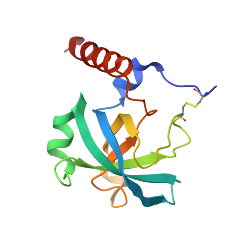
Structure and laminin-binding specificity of the NtA domain expressed in eukaryotic cells.
Mascarenhas, J.B., Ruegg, M.A., Sasaki, T., Eble, J.A., Engel, J., Stetefeld, J.(2005) Matrix Biol 23: 507-513
- PubMed: 15694127
- DOI: https://doi.org/10.1016/j.matbio.2004.11.003
- Primary Citation of Related Structures:
1PXU - PubMed Abstract:
Agrin is a key organizer for postsynaptic differentiation at the neuromuscular junction (NMJ). This activity requires the binding of agrin to the synaptic basal lamina via its N-terminal (NtA) domain. It has been suggested that this binding is mediated by conserved amino acids in the gamma 1 chain of laminin. Here, we report the crystal structure of chicken NtA expressed in eukaryotic HEK293 cells. In contrast to the previously published structure [Stetefeld, J., Jenny, M., Schulthess, T., Landwehr, R., Schumacher, B., Frank, S., Ruegg, M.A., Engel, J., Kammerer, R.A., 2001. The laminin-binding domain of agrin is structurally related to N-TIMP-1. Nat. Struct. Biol., 8, 705-709.], which was derived from the NtA domain expressed in E. coli, the new data show that the N-terminal tail region (amino acid residues Asn1-Arg5) is highly structured. Moreover, the disulfide bridge between Cys2 and Cys74 was also present. In addition, we show that the binding of NtA requires the gamma 1 chain of laminin and is not greatly affected by the composition of beta chains. These results confirm a model of the NtA-laminin complex where conserved amino acids in the gamma 1 chain are prerequisite for the binding to agrin and they further emphasize that the source of protein can be critical in structure determination.
Organizational Affiliation:
Biozentrum Universität Basel, Klingelbergstrasse 70, CH-4056 Basel, Switzerland.


