


Structure of an EngB GTPase
Kniewel, R., Buglino, J., Lima, C.D.To be published.
Experimental Data Snapshot
wwPDB Validation 3D Report Full Report

Entity ID: 1 | |||||
|---|---|---|---|---|---|
| Molecule | Chains | Sequence Length | Organism | Details | Image |
| Probable GTP-binding protein engB | 210 | Escherichia coli | Mutation(s): 0 Gene Names: ENGB | 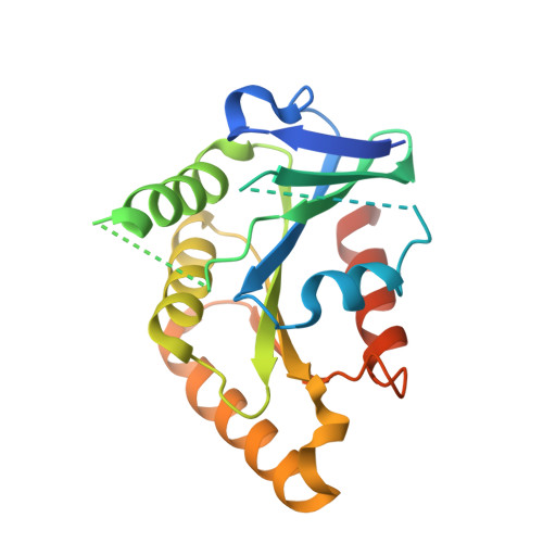 | |
UniProt | |||||
Find proteins for P0A6P7 (Escherichia coli (strain K12)) Explore P0A6P7 Go to UniProtKB: P0A6P7 | |||||
Entity Groups | |||||
| Sequence Clusters | 30% Identity50% Identity70% Identity90% Identity95% Identity100% Identity | ||||
| UniProt Group | P0A6P7 | ||||
Sequence AnnotationsExpand | |||||
| |||||
| Ligands 1 Unique | |||||
|---|---|---|---|---|---|
| ID | Chains | Name / Formula / InChI Key | 2D Diagram | 3D Interactions | |
| SO4 Query on SO4 | C [auth A] D [auth A] E [auth A] F [auth B] G [auth B] | SULFATE ION O4 S QAOWNCQODCNURD-UHFFFAOYSA-L |  | ||
| Length ( Å ) | Angle ( ˚ ) |
|---|---|
| a = 67.44 | α = 90 |
| b = 70.62 | β = 90 |
| c = 85.85 | γ = 90 |
| Software Name | Purpose |
|---|---|
| DENZO | data reduction |
| SCALEPACK | data scaling |
| SOLVE | phasing |
| RESOLVE | model building |
| CNS | refinement |
| RESOLVE | phasing |
RCSB PDB (citation) is hosted by
RCSB PDB is a member of the

