


1PTH
The Structural Basis of Aspirin Activity Inferred from the Crystal Structure of Inactivated Prostaglandin H2 Synthase
- PDB DOI: https://doi.org/10.2210/pdb1PTH/pdb
- Classification: OXIDOREDUCTASE
- Organism(s): Ovis aries
- Mutation(s): No
- Membrane Protein: Yes mpstruc
- Deposited: 1995-04-11 Released: 1996-04-11
Experimental Data Snapshot
- Method: X-RAY DIFFRACTION
- Resolution: 3.40 Å
- R-Value Free: 0.227
- R-Value Work: 0.186
- R-Value Observed: 0.186
wwPDB Validation 3D Report Full Report

This is version 2.0 of the entry. See complete history.

Macromolecules
Find similar proteins by:
(by identity cutoff) | 3D Structure
Entity ID: 1 | |||||
|---|---|---|---|---|---|
| Molecule | Chains | Sequence Length | Organism | Details | Image |
| PROSTAGLANDIN H2 SYNTHASE-1 | 576 | Ovis aries | Mutation(s): 0 EC: 1.14.99.1 Membrane Entity: Yes | 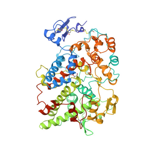 | |
UniProt | |||||
Find proteins for P05979 (Ovis aries) Explore P05979 Go to UniProtKB: P05979 | |||||
Entity Groups | |||||
| Sequence Clusters | 30% Identity50% Identity70% Identity90% Identity95% Identity100% Identity | ||||
| UniProt Group | P05979 | ||||
Sequence AnnotationsExpand | |||||
| |||||
Oligosaccharides

Small Molecules
| Ligands 4 Unique | |||||
|---|---|---|---|---|---|
| ID | Chains | Name / Formula / InChI Key | 2D Diagram | 3D Interactions | |
| HEM Query on HEM | H [auth A], M [auth B] | PROTOPORPHYRIN IX CONTAINING FE C34 H32 Fe N4 O4 KABFMIBPWCXCRK-RGGAHWMASA-L |  | ||
| BOG Query on BOG | G [auth A], L [auth B] | octyl beta-D-glucopyranoside C14 H28 O6 HEGSGKPQLMEBJL-RKQHYHRCSA-N |  | ||
| NAG Query on NAG | E [auth A], F [auth A], J [auth B], K [auth B] | 2-acetamido-2-deoxy-beta-D-glucopyranose C8 H15 N O6 OVRNDRQMDRJTHS-FMDGEEDCSA-N |  | ||
| SAL Query on SAL | I [auth A], N [auth B] | 2-HYDROXYBENZOIC ACID C7 H6 O3 YGSDEFSMJLZEOE-UHFFFAOYSA-N |  | ||
| Modified Residues 1 Unique | |||||
|---|---|---|---|---|---|
| ID | Chains | Type | Formula | 2D Diagram | Parent |
| 0AH Query on 0AH | A, B | L-PEPTIDE LINKING | C5 H8 Br N O4 |  | SER |
Experimental Data & Validation
Experimental Data
- Method: X-RAY DIFFRACTION
- Resolution: 3.40 Å
- R-Value Free: 0.227
- R-Value Work: 0.186
- R-Value Observed: 0.186
- Space Group: I 2 2 2
Unit Cell:
| Length ( Å ) | Angle ( ˚ ) |
|---|---|
| a = 99.57 | α = 90 |
| b = 209.8 | β = 90 |
| c = 235 | γ = 90 |
| Software Name | Purpose |
|---|---|
| MADNES | data collection |
| X-PLOR | model building |
| X-PLOR | refinement |
| MADNES | data reduction |
| X-PLOR | phasing |
Entry History
Deposition Data
- Released Date: 1996-04-11 Deposition Author(s): Loll, P.J., Picot, D., Garavito, R.M.
Revision History (Full details and data files)
- Version 1.0: 1996-04-11
Type: Initial release - Version 1.1: 2008-03-03
Changes: Version format compliance - Version 1.2: 2011-07-13
Changes: Non-polymer description, Version format compliance - Version 2.0: 2020-07-29
Type: Remediation
Reason: Carbohydrate remediation
Changes: Atomic model, Data collection, Derived calculations, Other, Structure summary
