Refined structure of the porin from Rhodopseudomonas blastica. Comparison with the porin from Rhodobacter capsulatus.
Kreusch, A., Schulz, G.E.(1994) J Mol Biol 243: 891-905
- PubMed: 7525973
- DOI: https://doi.org/10.1006/jmbi.1994.1690
- Primary Citation of Related Structures:
1PRN - PubMed Abstract:
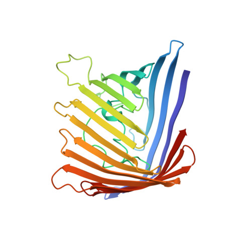
The structure of the membrane channel porin from the phototrophic bacteria Rhodopseudomonas blastica has been refined at 1.96 A resolution yielding an R-factor of 17.6%. The final model consists of all 289 amino acid residues, 247 water molecules and three detergent molecules modelled as n-octyltetraoxyethylene. One of these detergent molecules binds together with its two symmetry-related molecules tightly in a pocket at the molecular 3-fold axis. This pocket may bind three alkyl chains of a lipopolysaccharide which in turn would stabilize the trimer and could possibly play a role in membrane insertion. The overall shape of this porin resembles OmpF of Escherichia coli more than the only known sequence-related porin from Rhodobacter capsulatus. The membrane contacting surface is similar in all structurally known porins; it shows exceptional frequencies of amino acid residues and side-chain rotamers. The 46-residue loop beta 5-beta 6 of the porin is shown to be tightly fastened to the beta-barrel, excluding an in vivo loop movement that closes the pore. The trimer interface region has the structure of a water-soluble protein with an extensive non-polar core and numerous hydrogen bonds at the surface. The loops at the external end of the barrel are long and rigid whereas those at the periplasmic barrel end are short and mobile. The crystal packing is discussed.
Organizational Affiliation:
Institut für Organische Chemie und Biochemie, Albert-Ludwigs-Universität, Freiburg im Breisgau, Germany.