The refined crystal structure of dimeric phospholipase A2 at 2.5 A. Access to a shielded catalytic center.
Brunie, S., Bolin, J., Gewirth, D., Sigler, P.B.(1985) J Biol Chem 260: 9742-9749
- PubMed: 4019493
- Primary Citation of Related Structures:
1PP2 - PubMed Abstract:
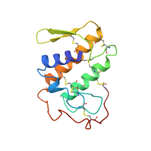
The 2.5-A crystal structure of the calcium-free form of the dimeric venom phospholipase A2 from the Western Diamondback rattlesnake Crotalus atrox, has been refined to an R-factor of 17.8% (I greater than 2 sigma) and acceptable stereochemistry. The molecule is a nearly perfect 2-fold symmetric dimer in which most of the catalytic residues of both subunits face an internal cavity. The restricted access to the putative catalytic sites is especially puzzling as the optimal substrates for this and most other phospholipase A2 are phospholipids condensed in micellar or lamellar aggregates. We point out that substrate access to the internal cavity may be aided by calcium binding which can alter the intersubunit contacts that shield the catalytic network. We also suggest that a system of hydrogen-bonded moieties exists on the surface of the dimer that links the amino terminus to the catalytic system, through an invariant Gln 4 side chain and the backbone of the active center residue, Tyr 73. This hydrogen-bonded network is on a highly accessible surface of the dimer and would appear to contribute to the enzyme's (as opposed to the proenzyme's) special capacity to attack aggregated rather than monomeric substrate.