Structure of the coat protein in Pf1 bacteriophage determined by solid-state NMR spectroscopy.
Thiriot, D.S., Nevzorov, A.A., Zagyanskiy, L., Wu, C.H., Opella, S.J.(2004) J Mol Biol 341: 869-879
- PubMed: 15288792
- DOI: https://doi.org/10.1016/j.jmb.2004.06.038
- Primary Citation of Related Structures:
1PJF - PubMed Abstract:
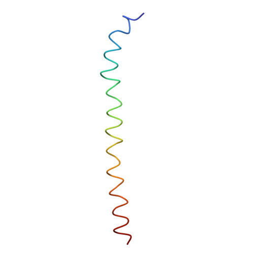
The atomic resolution structure of Pf1 coat protein determined by solid-state NMR spectroscopy of magnetically aligned filamentous bacteriophage particles in solution is compared to the structures previously determined by X-ray fiber and neutron diffraction, the structure of its membrane-bound form, and the structure of fd coat protein. These structural comparisons provide insights into several biological properties, differences between class I and class II filamentous bacteriophages, and the assembly process. The six N-terminal amino acid residues adopt an unusual "double hook" conformation on the outside of the bacteriophage particle. The solid-state NMR results indicate that at 30 degrees C, some of the coat protein subunits assume a single, fully structured conformation, and some have a few mobile residues that provide a break between two helical segments, in agreement with structural models from X-ray fiber and neutron diffraction, respectively. The atomic resolution structure determined by solid-state NMR for residues 7-14 and 18-46, which excludes the N-terminal double hook and the break between the helical segments, but encompasses more than 80% of the backbone including the distinct kink at residue 29, agrees with that determined by X-ray fiber diffraction with an RMSD value of 2.0 A. The symmetry and distance constraints determined by X-ray fiber and neutron diffraction enable the construction of an accurate model of the bacteriophage particle from the coordinates of the coat protein monomers.
Organizational Affiliation:
Department of Chemistry and Biochemistry, University of California, San Diego, CA 92093-0307, USA.