1OX3

crystal structure of mini-fibritin


Experimental Data Snapshot

  • Method: X-RAY DIFFRACTION
  • Resolution: 2.00 Å
  • R-Value Free: 0.221 
  • R-Value Work: 0.209 
  • R-Value Observed: 0.209 

wwPDB Validation   3D Report Full Report


This is version 1.5 of the entry. See complete history


Literature

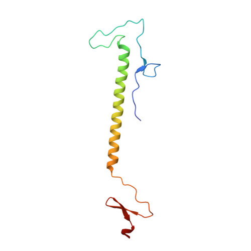
Design and Crystal Structure of Bacteriophage T4 Mini-Fibritin NCCF.

Boudko, S.P.Strelkov, S.V.Engel, J.Stetefeld, J.

(2004) J Mol Biol 339: 927-935

  • DOI: https://doi.org/10.1016/j.jmb.2004.04.001
  • Primary Citation of Related Structures:  
    1OX3

  • PubMed Abstract: 

    Fibritin is a fibrous protein that forms "whiskers" attached to the neck of bacteriophage T4. Whiskers interact with the long tail fibers regulating the assembly and infectivity of the virus. The fibritin trimer includes the N-terminal domain responsible for attachment to the phage particle and for the collar formation, the central domain forming a 500 A long segmented coiled-coil structure, and the C-terminal "foldon" domain. We have designed a "mini" fibritin with most of the coiled-coil domain deleted, and solved its crystal structure. The non-helical N-terminal part represents a new protein fold that tightly interacts with the coiled-coil segment forming a single domain, as revealed by calorimetry. The analysis of the crystal structure and earlier electron microscopy data on the collar-whisker complex suggests the necessity of other proteins to participate in the collar formation. Crystal structure determination of the N-terminal domain of fibritin is the first step towards elucidating the detailed structure and assembly mechanism of the collar-whisker complex.


  • Organizational Affiliation

    Biozentrum, University of Basel, Klingelbergstr.70, CH-4056 Basel, Switzerland. sergei.boudko@unibas.ch


Macromolecules
Find similar proteins by:  (by identity cutoff)  |  3D Structure
Entity ID: 1
MoleculeChains Sequence LengthOrganismDetailsImage
Fibritin109Tequatrovirus T4Mutation(s): 1 
Gene Names: wac
UniProt
Find proteins for P10104 (Enterobacteria phage T4)
Explore P10104 
Go to UniProtKB:  P10104
Entity Groups  
Sequence Clusters30% Identity50% Identity70% Identity90% Identity95% Identity100% Identity
UniProt GroupP10104
Sequence Annotations
Expand
  • Reference Sequence
Experimental Data & Validation

Experimental Data

  • Method: X-RAY DIFFRACTION
  • Resolution: 2.00 Å
  • R-Value Free: 0.221 
  • R-Value Work: 0.209 
  • R-Value Observed: 0.209 
  • Space Group: P 63
Unit Cell:
Length ( Å )Angle ( ˚ )
a = 54.615α = 90
b = 54.615β = 90
c = 107.848γ = 120
Software Package:
Software NamePurpose
CNSrefinement
MOSFLMdata reduction
CCP4data scaling
AMoREphasing

Structure Validation

View Full Validation Report


Entry History 

Deposition Data

Revision History  (Full details and data files)

  • Version 1.0: 2004-04-13
    Type: Initial release
  • Version 1.1: 2008-04-29
    Changes: Version format compliance
  • Version 1.2: 2011-07-13
    Changes: Derived calculations, Version format compliance
  • Version 1.3: 2011-11-16
    Changes: Atomic model
  • Version 1.4: 2017-06-28
    Changes: Advisory, Database references, Source and taxonomy, Structure summary
  • Version 1.5: 2023-08-16
    Changes: Data collection, Database references, Refinement description