Active site structural features for chemically modified forms of rhodanese.
Gliubich, F., Gazerro, M., Zanotti, G., Delbono, S., Bombieri, G., Berni, R.(1996) J Biol Chem 271: 21054-21061
- PubMed: 8702871
- DOI: https://doi.org/10.1074/jbc.271.35.21054
- Primary Citation of Related Structures:
1ORB, 2ORA - PubMed Abstract:
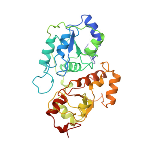
In the course of the reaction catalyzed by rhodanese, the enzyme cycles between two catalytic intermediates, the sulfur-free and the sulfur-substituted (persulfide-containing) forms. The crystal structure of sulfur-free rhodanese, which was prepared in solution and then crystallized, is highly similar to that of sulfur-substituted enzyme. The inactivation of sulfur-free rhodanese with a small molar excess of hydrogen peroxide relies essentially on a modification limited to the active site, consisting of the oxidation of the essential sulfhydryl to sulfenyl group (-S-OH). Upon reaction of the sulfur-free enzyme with monoiodoacetate in the crystal, the Cys-247 side chain with the bound carboxymethyl group is forced into a conformation that allows favorable interactions of the carboxylate with the four peptide NH groups that participate in hydrogen bonding interactions with the transferable sulfur atom of the persulfide group in the sulfur-substituted rhodanese. It is concluded that active site-specific chemical modifications of sulfur-free rhodanese do not lead to significant changes of the protein structure, consistent with a high degree of similarity of the structures of the sulfur-free and sulfur-substituted forms of the enzyme both in solution and in the crystal.
Organizational Affiliation:
Department of Organic Chemistry, University of Padova and Biopolymer Research Center, Consiglio Nazionale delle Ricerche, 35131 Padova, Italy.