Structure of the Neural (N-) Cadherin Prodomain Reveals a Cadherin Extracellular Domain-like Fold without Adhesive Characteristics
Koch, A.W., Farooq, A., Shan, W., Zeng, L., Colman, D.R., Zhou, M.-M.(2004) Structure 12: 793-805
- PubMed: 15130472
- DOI: https://doi.org/10.1016/j.str.2004.02.034
- Primary Citation of Related Structures:
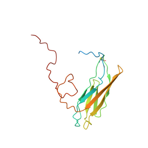
1OP4 - PubMed Abstract:
Classical cadherins mediate cell-cell adhesion through calcium-dependent homophilic interactions and are activated through cleavage of a prosequence in the late Golgi. We present here the first three-dimensional structure of a classical cadherin prosequence, solved by NMR. The prototypic prosequence of N-cadherin consists of an Ig-like domain and an unstructured C-terminal region. The folded part of the prosequence-termed prodomain-has a striking structural resemblance to cadherin "adhesive" domains that could not have been predicted from the amino acid sequence due to low sequence similarities. Our detailed structural and evolutionary analysis revealed that prodomains are distant relatives of cadherin "adhesive" domains but lack all the features known to be important for cadherin-cadherin interactions. The presence of an additional "nonadhesive" domain seems to make it impossible to engage homophilic interactions between cadherins that are necessary to activate adhesion, thus explaining the inactive state of prodomain-bearing cadherins.
Organizational Affiliation:
Fishberg Research Center for Neurobiology, Structural Biology Program, Department of Physiology and Biophysics, Mount Sinai School of Medicine, One Gustave L. Levy Place, New York, NY 10029 USA. akoch@gene.com