1OKJ

crystal structure of the essential E. coli YeaZ protein by MAD method using the gadolinium complex "DOTMA"


Experimental Data Snapshot

  • Method: X-RAY DIFFRACTION
  • Resolution: 2.28 Å
  • R-Value Free: 0.257 
  • R-Value Work: 0.216 
  • R-Value Observed: 0.216 

wwPDB Validation   3D Report Full Report


This is version 1.2 of the entry. See complete history


Literature

A Complement to the Modern Crystallographer'S Toolbox: Caged Gadolinium Complexes with Versatile Binding Modes.

Stelter, M.Molina, R.Jeudy, S.Kahn, R.Abergel, C.Hermoso, J.A.

(2014) Acta Crystallogr D Biol Crystallogr 70: 1506

  • DOI: https://doi.org/10.1107/S1399004714005483
  • Primary Citation of Related Structures:  
    1OKJ

  • PubMed Abstract: 

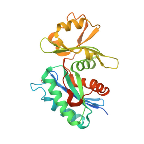
    A set of seven caged gadolinium complexes were used as vectors for introducing the chelated Gd(3+) ion into protein crystals in order to provide strong anomalous scattering for de novo phasing. The complexes contained multidentate ligand molecules with different functional groups to provide a panel of possible interactions with the protein. An exhaustive crystallographic analysis showed them to be nondisruptive to the diffraction quality of the prepared derivative crystals, and as many as 50% of the derivatives allowed the determination of accurate phases, leading to high-quality experimental electron-density maps. At least two successful derivatives were identified for all tested proteins. Structure refinement showed that the complexes bind to the protein surface or solvent-accessible cavities, involving hydrogen bonds, electrostatic and CH-π interactions, explaining their versatile binding modes. Their high phasing power, complementary binding modes and ease of use make them highly suitable as a heavy-atom screen for high-throughput de novo structure determination, in combination with the SAD method. They can also provide a reliable tool for the development of new methods such as serial femtosecond crystallography.


  • Organizational Affiliation

    University Grenoble Alpes, Institut de Biologie Structurale (IBS), Grenoble, France.


Macromolecules
Find similar proteins by:  (by identity cutoff)  |  3D Structure
Entity ID: 1
MoleculeChains Sequence LengthOrganismDetailsImage
TRNA THREONYLCARBAMOYLADENOSINE BIOSYNTHESIS PROTEIN TSAB
A, B, C, D
251Escherichia coli K-12Mutation(s): 0 
EC: 3.4
UniProt
Find proteins for P76256 (Escherichia coli (strain K12))
Explore P76256 
Go to UniProtKB:  P76256
Entity Groups  
Sequence Clusters30% Identity50% Identity70% Identity90% Identity95% Identity100% Identity
UniProt GroupP76256
Sequence Annotations
Expand
  • Reference Sequence
Experimental Data & Validation

Experimental Data

  • Method: X-RAY DIFFRACTION
  • Resolution: 2.28 Å
  • R-Value Free: 0.257 
  • R-Value Work: 0.216 
  • R-Value Observed: 0.216 
  • Space Group: P 21 21 21
Unit Cell:
Length ( Å )Angle ( ˚ )
a = 76.34α = 90
b = 97.6β = 90
c = 141.94γ = 90
Software Package:
Software NamePurpose
MOSFLMdata reduction
SCALAdata scaling
SOLVEphasing
autoSHARPphasing
CNSrefinement

Structure Validation

View Full Validation Report


Entry History 

Deposition Data

Revision History  (Full details and data files)

  • Version 1.0: 2004-09-16
    Type: Initial release
  • Version 1.1: 2014-02-05
    Changes: Database references, Derived calculations, Non-polymer description, Other, Refinement description, Source and taxonomy, Structure summary, Version format compliance
  • Version 1.2: 2014-07-23
    Changes: Database references, Other, Structure summary