1OAE

Crystal structure of the reduced form of cytochrome c" from Methylophilus methylotrophus


Experimental Data Snapshot

  • Method: X-RAY DIFFRACTION
  • Resolution: 1.95 Å
  • R-Value Free: 0.243 
  • R-Value Work: 0.185 
  • R-Value Observed: 0.188 

wwPDB Validation   3D Report Full Report


Ligand Structure Quality Assessment 


This is version 1.2 of the entry. See complete history


Literature

Structural Evidence for a Proton Transfer Pathway Coupled with Haem Reduction of Cytochrome C" from Methylophilus Methylotrophus.

Enguita, F.J.Pohl, E.Turner, D.L.Santos, H.Carrondo, M.A.

(2006) J Biol Inorg Chem 11: 189

  • DOI: https://doi.org/10.1007/s00775-005-0065-6
  • Primary Citation of Related Structures:  
    1GU2, 1OAE

  • PubMed Abstract: 

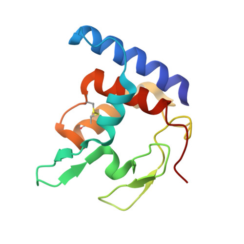
    The crystal structures of the oxidized and reduced forms of cytochrome c" from Methylophilus methylotrophus were solved from X-ray synchrotron data to atomic resolution. The overall fold of the molecule in the two redox states is very similar and is comparable to that of the oxygen-binding protein from the purple phototrophic bacterium Rhodobacter sphaeroides. However, significant modifications occur near the haem group, in particular the detachment from axial binding of His95 observed upon reduction as well as the adoption of different conformations of some protonatable residues that form a possible proton path from the haem pocket to the protein surface. These changes are associated with the previously well characterized redox-Bohr behaviour of this protein. Furthermore they provide a model for one of the presently proposed mechanisms of proton translocation in the much more complex protein cytochrome c oxidase.


  • Organizational Affiliation

    Instituto de Tecnologia Química e Biológica, Universidade Nova de Lisboa, P.O. Box 127, 2781-901 Oeiras, Portugal.


Macromolecules
Find similar proteins by:  (by identity cutoff)  |  3D Structure
Entity ID: 1
MoleculeChains Sequence LengthOrganismDetailsImage
CYTOCHROME C"
A, B
124Methylophilus methylotrophusMutation(s): 0 
UniProt
Find proteins for Q9RQB9 (Methylophilus methylotrophus)
Explore Q9RQB9 
Go to UniProtKB:  Q9RQB9
Entity Groups  
Sequence Clusters30% Identity50% Identity70% Identity90% Identity95% Identity100% Identity
UniProt GroupQ9RQB9
Sequence Annotations
Expand
  • Reference Sequence
Small Molecules
Ligands 3 Unique
IDChains Name / Formula / InChI Key2D Diagram3D Interactions
HEC
Query on HEC

Download Ideal Coordinates CCD File 
C [auth A],
J [auth B]
HEME C
C34 H34 Fe N4 O4
HXQIYSLZKNYNMH-LJNAALQVSA-N
SO4
Query on SO4

Download Ideal Coordinates CCD File 
G [auth A]
H [auth A]
I [auth A]
M [auth B]
N [auth B]
G [auth A],
H [auth A],
I [auth A],
M [auth B],
N [auth B],
O [auth B]
SULFATE ION
O4 S
QAOWNCQODCNURD-UHFFFAOYSA-L
GOL
Query on GOL

Download Ideal Coordinates CCD File 
D [auth A],
E [auth A],
F [auth A],
K [auth B],
L [auth B]
GLYCEROL
C3 H8 O3
PEDCQBHIVMGVHV-UHFFFAOYSA-N
Experimental Data & Validation

Experimental Data

  • Method: X-RAY DIFFRACTION
  • Resolution: 1.95 Å
  • R-Value Free: 0.243 
  • R-Value Work: 0.185 
  • R-Value Observed: 0.188 
  • Space Group: P 21 21 21
Unit Cell:
Length ( Å )Angle ( ˚ )
a = 58.688α = 90
b = 74.362β = 90
c = 77.842γ = 90
Software Package:
Software NamePurpose
REFMACrefinement
MOSFLMdata reduction
SCALAdata scaling
MOLREPphasing

Structure Validation

View Full Validation Report


Ligand Structure Quality Assessment 


Entry History 

Deposition Data

Revision History  (Full details and data files)

  • Version 1.0: 2004-03-26
    Type: Initial release
  • Version 1.1: 2014-10-22
    Changes: Database references, Derived calculations, Other, Version format compliance
  • Version 1.2: 2019-07-24
    Changes: Data collection, Derived calculations