1O57

CRYSTAL STRUCTURE OF THE PURINE OPERON REPRESSOR OF BACILLUS SUBTILIS


Experimental Data Snapshot

  • Method: X-RAY DIFFRACTION
  • Resolution: 2.20 Å
  • R-Value Free: 0.237 
  • R-Value Work: 0.188 
  • R-Value Observed: 0.100 

wwPDB Validation   3D Report Full Report


This is version 1.3 of the entry. See complete history


Literature

The Purine Repressor of Bacillus Subtilis: A Novel Combination of Domains Adapted for Transcription Regulation

Sinha, S.C.Krahn, J.Shin, B.S.Tomchick, D.R.Zalkin, H.Smith, J.L.

(2003) J Bacteriol 185: 4087-4098

  • DOI: https://doi.org/10.1128/JB.185.14.4087-4098.2003
  • Primary Citation of Related Structures:  
    1O57

  • PubMed Abstract: 

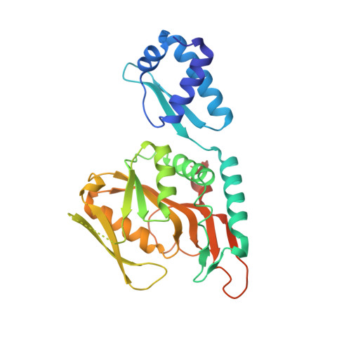
    The purine repressor from Bacillus subtilis, PurR, represses transcription from a number of genes with functions in the synthesis, transport, and metabolism of purines. The 2.2-A crystal structure of PurR reveals a two-domain protein organized as a dimer. The larger C-terminal domain belongs to the PRT structural family, in accord with a sequence motif for binding the inducer phosphoribosylpyrophosphate (PRPP). The PRT domain is fused to a smaller N-terminal domain that belongs to the winged-helix family of DNA binding proteins. A positively charged surface on the winged-helix domain likely binds specific DNA sequences in the recognition site. A second positively charged surface surrounds the PRPP site at the opposite end of the PurR dimer. Conserved amino acids in the sequences of PurR homologs in 21 gram-positive bacteria cluster on the proposed recognition surface of the winged-helix domain and around the PRPP binding site at the opposite end of the molecule, supporting a common function of DNA and PRPP binding for all of the proteins. The structure supports a binding mechanism in which extended regions of DNA interact with extensive protein surface. Unlike most PRT proteins, which are phosphoribosyltransferases (PRTases), PurR lacks catalytic activity. This is explained by a tyrosine side chain that blocks the site for a nucleophile cosubstrate in PRTases. Thus, B. subtilis has adapted an enzyme fold to serve as an effector-binding domain and has used it in a novel combination with the DNA-binding winged-helix domain as a repressor of purine genes.


  • Organizational Affiliation

    Department of Biological Sciences, Purdue University, West Lafayette, Indiana 47907, USA.


Macromolecules
Find similar proteins by:  (by identity cutoff)  |  3D Structure
Entity ID: 1
MoleculeChains Sequence LengthOrganismDetailsImage
PUR OPERON REPRESSOR
A, B, C, D
291Bacillus subtilisMutation(s): 0 
Gene Names: PURR
UniProt
Find proteins for P37551 (Bacillus subtilis (strain 168))
Explore P37551 
Go to UniProtKB:  P37551
Entity Groups  
Sequence Clusters30% Identity50% Identity70% Identity90% Identity95% Identity100% Identity
UniProt GroupP37551
Sequence Annotations
Expand
  • Reference Sequence
Small Molecules
Ligands 6 Unique
IDChains Name / Formula / InChI Key2D Diagram3D Interactions
2PE
Query on 2PE

Download Ideal Coordinates CCD File 
H [auth A]NONAETHYLENE GLYCOL
C18 H38 O10
YZUUTMGDONTGTN-UHFFFAOYSA-N
P6G
Query on P6G

Download Ideal Coordinates CCD File 
G [auth A],
S [auth D]
HEXAETHYLENE GLYCOL
C12 H26 O7
IIRDTKBZINWQAW-UHFFFAOYSA-N
EPE
Query on EPE

Download Ideal Coordinates CCD File 
F [auth A],
R [auth D]
4-(2-HYDROXYETHYL)-1-PIPERAZINE ETHANESULFONIC ACID
C8 H18 N2 O4 S
JKMHFZQWWAIEOD-UHFFFAOYSA-N
1PE
Query on 1PE

Download Ideal Coordinates CCD File 
P [auth C],
T [auth D]
PENTAETHYLENE GLYCOL
C10 H22 O6
JLFNLZLINWHATN-UHFFFAOYSA-N
PG4
Query on PG4

Download Ideal Coordinates CCD File 
L [auth B]TETRAETHYLENE GLYCOL
C8 H18 O5
UWHCKJMYHZGTIT-UHFFFAOYSA-N
SO4
Query on SO4

Download Ideal Coordinates CCD File 
E [auth A]
I [auth B]
J [auth B]
K [auth B]
M [auth C]
E [auth A],
I [auth B],
J [auth B],
K [auth B],
M [auth C],
N [auth C],
O [auth C],
Q [auth D]
SULFATE ION
O4 S
QAOWNCQODCNURD-UHFFFAOYSA-L
Experimental Data & Validation

Experimental Data

  • Method: X-RAY DIFFRACTION
  • Resolution: 2.20 Å
  • R-Value Free: 0.237 
  • R-Value Work: 0.188 
  • R-Value Observed: 0.100 
  • Space Group: P 1
Unit Cell:
Length ( Å )Angle ( ˚ )
a = 65.077α = 84.75
b = 72.156β = 84.03
c = 82.967γ = 67.47
Software Package:
Software NamePurpose
DENZOdata reduction
SCALEPACKdata scaling
MLPHAREphasing
CNSrefinement

Structure Validation

View Full Validation Report


Entry History 

Deposition Data

Revision History  (Full details and data files)

  • Version 1.0: 2003-08-26
    Type: Initial release
  • Version 1.1: 2008-04-26
    Changes: Version format compliance
  • Version 1.2: 2011-07-13
    Changes: Version format compliance
  • Version 1.3: 2023-12-27
    Changes: Data collection, Database references, Derived calculations